Bridging Two Creative Universes - Science Art Show

This exhibition explores the fascinating intersection of science and art bringing together the works of talented researchers and artists who have been inspired by the wonders of biology. This interactive exhibit exemplifies how art can communicate complex scientific concepts, foster innovative collaborations, and advance scientific discovery. Scientists and artists put forward a diverse range of art pieces, including paintings, sculptures, fashion, and interactive media that showcase the powerful connection between science and art, emphasizing how artistic practices are transforming scientific research, communication, and visualization through creativity and cutting-edge technologies.

Photos

Group photo of in-person exhibiting artists

Photo gallery

Exhibiting Artists/Scientists

Digital Exhibit

Joshua Almond

Saddleback College; MiraCosta College

Joshua Almond is an artist/craftsman living and working in Orange County, California. Joshua works primarily in wood to create abstract landscapes, studio furniture, and large-scale sculptural forms that explore the sublime beauty inherent in microbiological/cellular structures. Born and raised in Southern New England, Joshua received his BA from Rhodes College and his MFA from the Herberger Institute for Design and the Arts: School of Art at Arizona State University.

Things: COS-7 No. 4 (2023)

Artist: Joshua Almond

Graphite on paper

Series of drawings derived from studies of COS-7 cells.

Please contact the artist at www.joshalmond.com for information on usage and permissions.

Covid Stool (2021)

Artist: Joshua Almond

White oak

Shikaku-style stake leg footstool with Covid-19 motif.

Please contact the artist at www.joshalmond.com for information on usage and permissions.

Oak Leaf Stool (2021)

Artist: Joshua Almond

White oak

Shikaku-style stake leg footstool with cellular-level oak leaf motif.

Please contact the artist at www.joshalmond.com for information on usage and permissions.

Hugo Heredia Barrera

My name is Hugo Heredia Barrera and I am an artist based in San Diego, CA. I have vast experience working with many different types of art, from exhibitions in museums, fine art galleries, public fine art fairs, and public art projects. In my work I incorporate diverse techniques and ideas, from simple mechanical sculptures to organic and architectural art installations. Most of my art and art projects integrate technology elements as I make art with different glass working techniques and different metal and metal working techniques. I have spent most of my art career experimenting with glass for scientific purposes and I have worked on glass projects for companies including NASA, Boeing Corporation and Lockheed Martin. Most recently, I alternate my art career with aerospace as I have worked with companies such as General Atomics, Virgin Orbit and SpaceX.

Among some of my most recent art projects are an installation for a show called “Illumination: 21st Century Interactions with Art, Science and Technology” at the San Diego Art Institute in 2020. Here, 25 artists were paired with 25 scientists, with each team presenting a different sculpture or installation that combined art and science. For my installation, I was inspired by the research of a scientist working for UC San Diego to create an immersive sculpture installation where I explored how cancer spreads within the body. With this project I not only explore technology for the purpose of fusing and shaping the glass, but also the artwork is used to communicate scientific concepts to the viewer. Besides this installation, I had an Immersive installation show at San Diego City College, and at The International San Diego Airport in which both of them I used some kind of technology.

Sueño Rosa (2015)

Artist: Hugo Heredia Barrera

Cast glass, fabricated steel with bronze

Casted glass, torch cut steel welded with bronze and patina.

Sueño Rosa is inspired by the beautiful, fascinating and intricate process of embryonic development and the life cycle of an organism. The rounded glass is reminiscent of a fertilized egg in its early stages and the rose tainted circles resemble the cells of the embryo. Living beings are defined by opposing features: fragile and vulnerable, but also strong and resilient. These contradictions are represented by the mixture between two different types of materials: a delicate piece of glass and a rigid, spiral shaped metal.

Please contact Hugo Heredia Barrera for information on usage and permissions

Drew Berry

WEHI Medical Research, Melbourne Australia

wehi.tv

Drew Berry is a biologist-animator renowned for his visually stunning, scientifically accurate animations of molecular and cellular processes. Beginning his career as a cell biologist, Drew is fluent in navigating technical reports and research data from scientific journals. As an artist, he transforms complicated scientific concepts into vivid and meaningful animated journeys.  Since 1995, Drew has led biomedical animation within WEHI (Melbourne), Australia’s flagship for medical research. His work has showcased at the Guggenheim Museum, MoMA, V&A and the Royal Institute of Great Britain. In 2011, he collaborated with Björk on her app album and music video for the ‘Biophilia’ tour. Drew has received numerous awards, including an Emmy, a BAFTA, and the MacArthur Fellowship “Genius Grant”.

DNA Break Repair by Homologous Recombination (2024)

Artist: Drew Berry

Biomolecular Animation

Homologous recombination is crucial in repairing double-strand breaks in DNA and maintaining genomic integrity. 

Meticulously reconstructed using molecular structures from 3D cryo-EM tomography, molecular dynamics simulation, and AlphaFold, this animation unveils the intricate molecular mechanism your cells employ to repair double-strand DNA break damage without errors. It’s a powerful educational tool that informs and empowers you with a deeper understanding of this complex process. 

This animation brings into focus the indispensable role of BRCA1 and BRCA2 in DNA break repair in homologous recombination. It’s designed to support outreach on WEHI’s breast cancer research, connecting you to these key players in the fight against breast cancer and providing world-class education materials on foundational cell biology topics.

Freely available for use under a CC-BY-4.0 license.

Laura Olivares Boldú

Wellcome Connecting Science

Laura is a visual communicator of science at Wellcome Connecting Science in Cambridge (UK). She creates illustrations, animations, data visualisations and graphic materials that promote the understanding and exploration of genomic science and its impact.

Laura has a background in Biotechnology and Biomedical research, and over ten years of experience working as a graphic designer and illustrator in Cambridge, London and Barcelona. She is passionate about taking a complex concept and transforming it into something visual that enables people to engage with it.

Prime Editing (2023)

Artist: Laura Olivares Boldú

Digital

Illustration showing the components of the prime editing system and how it works. Designed for an explainer page on the prime editing technique.

Freely available for use under a CC BY-NC-SA 4.0 license. Please, credit the work to ‘Laura Olivares Boldu / Wellcome Connecting Science’.

Conversation on the Return of Additional Findings From Genetic Research (2023)

Artist: Laura Olivares Boldú

Digital

Animated illustration for the event ‘IBD BioResource genetic feedback: a conversation between participants and researchers’. The workshop aimed to create an open conversation between researchers, clinicians, and IBD BioResource participants on how to return additional findings in genomics research. The illustration consist of a gut with a speech bubble on top that reveals double strands of DNA. There is a DNA section highlighted in orange, conveying the ‘additional findings’ discussed at the workshop.

Freely available for use under a CC BY-NC-SA 4.0 license. Please credit the work to ‘Laura Olivares Boldu / Wellcome Connecting Science’.

Patricia Bondia

Scientific Videogames

I’m Patricia Bondia, a scientist turned visual communicator. During my PhD researching biology at the nanoscale, I discovered how fascinating science can be, but also how challenging it is to communicate it in a way that truly engages others. Motivated by a desire to improve effective communication, I began creating illustrations designed to spark curiosity and encourage deeper exploration. Through this process, I realized the power of visuals in capturing attention and inspiring engagement. Eager to push this further, I co-founded Scientific Videogames to transform visuals into immersive and interactive experiences. Today, I collaborate with research centers and companies worldwide to transform complex ideas into captivating visuals, animations, and interactive adventures. I believe that engaging communication is the key to sparking scientific vocations in future generations.

Light-Induced Phase Transitions (2023)

Artist: Patricia Bondia

Digital

The illustration depicts a crystalline lattice melting (represented as a snowflake), superimposed with its coherent X-ray scattering pattern. It shows light inducing a change state from solid to liquid.

It is a visualization inspired by the research of Simon Wall and co-workers: Johnson, A.S., Perez-Salinas, D., Siddiqui, K.M. et al. Ultrafast X-ray imaging of the light-induced phase transition in VO2. Nat. Phys. 19, 215–220 (2023).

https://doi.org/10.1038/s41567-022-01848-w

Please contact Patricia Bondia for information on usage and permissions.

Nanohispa Car (2023)

Artist: Patricia Bondia

Digital

This image showcases Nanohispa, the winning nanocar of the Nanocar Race, an epic competition where molecules speed across a track made of gold atoms. To celebrate the victory of the nanocar designed by IMDEA Nanoscience, my associate Melek Villanueva and I collaborated with them to create Nanocar Racing: The Game—a fun, fast-paced, and interactive way to experience the thrill of molecular racing. Ready to take on the challenge? Play it now on PC, Android, and iPhone, and see if you can dominate the nanoscale track!

Please contact Patricia Bondia for information on usage and permissions.

Perovskite Solar Cell (2023)

Artist: Patricia Bondia

Digital

This animation illustrates the components of a solar cell, highlighting its key feature: the use of molecular switches, molecules that react to light. The secret to its efficiency lies in strategically positioning these molecules between layers of perovskite structures. As the animation zooms out, we see how it integrates into the complete device. This project was commissioned by Agata Communication.

Please contact Patricia Bondia for information on usage and permissions.

Chiral Materials Boost Light Capture for Improved Hydrogen Production (2023)

Artist: Patricia Bondia

Digital

This cover illustrates a breakthrough in hydrogen production using cellulose nanocrystals to build photoactive black titania. The study offers insights into designing nanostructured materials to enhance light harvesting, unlocking new possibilities for renewable energy through sunlight. The research was led by Mohamed Nawfal Ghazzal and his team (Institut de Chimie Physique-CNRS).

Please contact Patricia Bondia for information on usage and permissions.

Timothy Davison

CellWalk

Tim Davison is a scientist and innovator transforming biology education through immersive and interactive storytelling. As the creator of the CellWalk project, he enables learners to explore entire cells—from large-scale structures down to individual atoms—through interactive and immersive visualization on everyday devices. Apple has featured CellWalk on both the iOS and Apple Vision Pro App Stores.

Tim earned his PhD in computer graphics, specializing in interactive and immersive visualization of mesoscale scenes using spatial computing. Driven by a passion for user experience and design, he believes that beautiful, accurate visuals are the best ways to engage learners and inspire excitement for science. By pushing the boundaries of interactive learning, he’s helping to shape the future of biology education.

Exploring a Cell (2024)

Artist: Tim Davison

Exploring a dense and chaotic bacteria cell. Immersive and interactive tools bring us inside scientific data to make it accessible and engaging. Beautiful visualization and art is one of the best tools we have to connect learners with incredible science.

CC-BY-4.0 license

Suddenly a Wild Mycoplasma Appears (iPhone AR) (2024)

Artist: Tim Davison

Spatial computing technologies allow us to merge the real world with the microscopic one. Here, a tiny microscopic bacteria becomes a part of our world, for us to explore and connect with. 

CC-BY-4.0 license


Thao Do

Allen Institute for Cell Science; Do.T Studio

Thao Do is a user experience designer specializing in science applications, and also a science communicator with a diverse range of creative interests & skills in illustration, animation, graphic design, comics, and crafts. Growing up in southern Vietnam, she spent time in her grandparents’ expansive orchard, her father’s beautiful orchid garden and serene fish ponds, all set on fertile lands and surrounded by rich alluvial rivers. This experience has inspired her imagination and provoked her curiosity for the natural world, which eventually led her to pursue both science and art fields in her career development and became who she is today.

Dorotea Fracchiolla - Art&Science

Scientific Illustrator at Art&Science; Project Manager at Max Planck Institute for Biophysics, Frankfurt am Main

Dorotea Fracchiolla is an Italian-born scientist with a lifelong passion for visual arts and communication. Born in Bari she is currently Project Manager of mito911, an international and multi-institutional team studying mitophagy in Parkinson’s disease and a freelancer scientific illustrator and science communicator. She holds a PhD in Molecular Biology from the University of Vienna (AT) where she worked with Prof. Sascha Martens on selective autophagy in the budding yeast. The in vitro reductionist approach used in the lab inspired her art. Dorotea works by the motto “if you can draw it, you understand it to the very detail”. Her style, inspired by the colorful landscapes of her land of origin Puglia, soon became appreciated by the general public who more easily was introduced to specialized knowledge via her appealing artworks. Her work appeared on covers of the Autophagy, Molecular Cell, Cell Journals and continues to inspire professionals and the general public who through art more easily get introduced to science.

Rising of Parkinson’s Disease (2023)

Dorotea Fracchiolla - Art&Science

Acrylics on canvas

This painting is part of a collection, the Parkinson’s Disease (PD) Edition and shows the research lines on PD carried out within the Collaborative Research Network of ASAP, in which Dorotea works. The trees stemming from the ground represent the brain as it progresses through disease when going from day to night. Impairment of dopaminergic neurons, the cells primarily affected in PD, is represented as red apples growing at the basis of the tree crowns, corresponding to an area of the brain called substantia nigra where these cells are localized. Deep in the soil, the plant roots mimic the highly arborised dopaminergic neuron terminals, as they appear inside the brain. With disease progression, dopaminergic neurons lose the ability to secrete dopamine (shown as white speckles), an excitatory neurotransmitter that mediates their function. In an opposite trend, pathological inclusions (shown as dark stones) start to accumulate in these neurons and in the space around them affecting their function. The water streams penetrating the soil represent the attempt of providing therapies that would sustain dopaminergic neuronal activity and ameliorate disease. To achieve this, basic research stays central: on the left side of the planet, two cellular processes crucial in PD pathogenesis are illustrated, the degradation of mitochondria, energy factories of the cell, also known as mitophagy (at the Gulf of Mexico), and the endo-lysosomal trafficking pathway (in the Atlantic Ocean). Research in ASAP is also conducted to study brain circuit functionalities in whole animal systems, such as non-human primates. The painting celebrates all members of the ASAP network, visible as silhouettes at the horizon and travelling to San Diego city where the 2023 ASAP meeting took place, following which this painting was created.

Please contact Dorotea Fracchiolla for information on usage and permissions.

Same Meal, Different Tools - Mitophagy (2023)

Dorotea Fracchiolla - Art&Science

Photograph

In cells, damaged mitochondria are engulfed within a double membrane sac called “autophagosome” and selectively removed for degradation by the autophagy machinery to restrict cellular damage, a process called mitophagy. In neuronal cells, failure to do so is linked to neurodegenerative diseases such as Parkinson’s Disease. In Nguyen et al. Mol Cell (2023), authors uncover an unconventional mechanism for the selective removal of mitochondria upon induction of PINK1/Parkin signalling. They show that the cargo receptor Optineurin (OPTN) functions distinctly from the well known cargo receptor Nuclear Dot Protein 52 (NDP52). The artwork shows a roasted potato (the damaged mitochondria) being picked up from the plate by two different sets of table cutlery: chopsticks or fork and knife (that is OPTN or NDP52). The potato will be eaten up by the diners in either case, but picked up in two different ways!

Please contact Dorotea Fracchiolla for information on usage and permissions.

Coronavirus Goes on a Journey (2020)

Dorotea Fracchiolla - Art&Science

Gouache and ink on paper

This illustration was made to present Coronavirus to the general public. The author recreated the moment in which the virus prepares its luggage before going on a journey around the world. Every element shown in the image metaphorically represents the function of the proteins encoded in the viral RNA (orange rod inside the luggage in the middle of the illustration). Viruses are not living organisms but when they find a suitable host they can exploit its machinery to replicate, survive and propagate. A ticket with the Spike protein drawn on it represents what the virus uses to enter the host cell. There are also a lot of scissors: they represent Endonucleases, the enzymes used to cleave the host cell DNA. A pair of glasses and moustache placed on viral RNA represent the tools used by the virus to hide and sabotage the host cell. Now, try to guess what the other items in the picture represent or ask Dorotea to reveal more about them!

Please contact Dorotea Fracchiolla for information on usage and permissions.

A Graceful Ballet of Molecules (2020)

Dorotea Fracchiolla - Art&Science

Colored pencils and ink on paper

This illustration represents the ATG8 lipidation to Phosphatidylethanolamine reaction during autophagosome formation. Like in a graceful ballet, one after the other each molecule comes in place on the autophagosomal membrane to perform its own task. In Fracchiolla et al. JCB (2020) authors mechanistically dissect each step of this chain of reactions and reconstitute the entire process in vitro using recombinant proteins and synthetic membranes. This illustration was used as Graphical Abstract of the linked research article.

Please contact Dorotea Fracchiolla for information on usage and permissions.

Mosaic Cortical Neuron Organoid as a Tool to Study Population Genetics (2024)

Artist: Dorotea Fracchiolla - Art&Science

Digital illustration

Caporale, N., Castaldi, D., Rigoli, M.T. et al. develop a method to multiplex cortical brain organoids for the longitudinal dissection of developmental traits at single-cell resolution. The illustration shows how pluripotent stem cells (single balloons) from different donors/individuals can be used by researchers to grow mosaic organoids in vitro (hot-air balloon) opening the possibility to use this tool for the study of population (shown as silhouette outlines) genetics. Shadows on the ground show how the organoid mimics the organ of the brain and that it can be used to follow single cells developmental trajectories.

Please contact Dorotea Fracchiolla for information on usage and permissions.

Uncovering the Marvels of a Synovial Myeloid Tissue Dendritic Cells Mosaic (2024)

Dorotea Fracchiolla - Art&Science

Digital illustration

Dendritic cell (DC) regulation of human tissue immunity remains unclear. MacDonald et al. investigate the roles of synovial tissue (ST) myeloid DCs in health and active or remission stages of rheumatoid arthritis. Using a combination of scRNAseq, spatial transcriptomics and DC/T cell co-cultures, the authors reveal functional features of ST-DCs that infer their roles in immune tolerance and disease. The illustration shows a team of archeologists unravelling the pattern of a mosaic floor in an ancient domus romana. The colorful tiles pattern represents the newly characterised composition and localization of diverse DC/T cell populations found in the synovial tissue (mosaic floor). Below, the hypocaustum represents the underlying blood stream from where DCs infiltrate the synovial tissue (holes in the floor and fallen mosaic pieces show connection between the two levels) determining the three conditions analysed. This illustration was chosen as the cover of the Immunity Cell Journal, Number 57, Issue 12.

Please contact Dorotea Fracchiolla for information on usage and permissions.

Gloria Fuentes

Visual Thinker

Gloria has a background in Organic and Physical Chemistry and holds a Ph.D. in Molecular Biology and Enzymology. Throughout her scientific journey, she’s spent most of her career exploring the exciting overlap between Structural Computational Biology and Biomedicine. Her adventures as a researcher have taken her to wonderful places like the UK, The Netherlands, Spain, Singapore, and Japan, enriching her understanding of both science and life in truly unique ways. 

After becoming a mother, she decided it was time for a change in her career path and beautifully transitioned into the world of illustration and animation. This new journey allows her to blend her love for science with her artistic passions. Driven by a desire to enhance scientific communication between her fellow researchers and the broader community, she established a small studio called Visual Thinker. Her inspiring motto is: “Let’s unleash creativity to bridge the gap between science and society. If I can illustrate it, you can understand it.”

Plectasin's Supramolecular Killing Mechanism (2024)

Artist: Gloria Fuentes - Visual Thinker

Digital

This illustration shows how plectasin, a fungal antibiotic studied by Markus Weingarth’s group at Utrecht University, acts by forming dense structures on bacterial membranes with lipid II through a calcium-sensitive mechanism. It highlights the oligomerization of plectasin molecules into larger complexes, facilitated by calcium binding to its anionic patch. This arrangement is vital for its effectiveness in disrupting bacterial cell wall synthesis, offering a new strategy against antimicrobial resistance.

Please contact gloglita@thevisualthinker.xyz for information on usage and permissions.

Different Therapies at Various Molecular Levels To Treat Diseases: Too Many Heads for One Hat (2019)

Artist: Gloria Fuentes - Visual Thinker

Digital

This infographic offers a detailed overview of various therapeutic strategies implemented at different molecular levels for disease treatment. It ranges from CRISPR-Cas9 techniques that target DNA strands to the use of genetically engineered therapeutic antibodies that attach to cell surfaces. Additionally, it includes antisense oligonucleotides—synthetic nucleic acid sequences that bind to specific RNA strands—RNA interference with double-stranded RNA that interrupts gene expression, and small molecule drugs that interact with proteins. This wide array of therapeutic tools emphasizes the idea that multiple solutions exist for a single issue.

Freely available for use under a CC-BY-4.0 license.

Exploring the Rich Diversity of Asian Genetics: A Journey through Singapore's Genomic Tapestry (2019)

Artist: Gloria Fuentes - Visual Thinker

Digital

Asian genetic diversity is underrepresented in genomics research, which hampers understanding of population structure, disease-related variants, and precision medicine in Asia. Wu et al. (736–749) conduct a whole-genome analysis of Chinese, Malay, and Indian genomes in Singapore, identifying novel Asian-specific variants, uncovering demographic histories, and finding disease variants enriched in natural selection regions. This diversity improves genetic data inference accuracy for other Asian and Oceanian populations. The cover depicts genetic diversity in Singapore, featuring individuals from major ethnic groups around a Singaporean flag with a DNA sequence.

Please contact gloglita@thevisualthinker.xyz for information on usage and permissions.

Jaye Gardiner

Fox Chase Cancer Center; JKX Comics

For as long as she can remember, Dr. Jaye Gardiner (she/her) has been interested in both science and art. First wowed by science after placing celery in colored water in her Chicago classroom, she’d also spend her spare time learning to draw her favorite cartoon characters. She originally assumed she’d have to choose between passions, but graduating from Macalester College with her Bachelor of Arts degree showed her the value of interdisciplinary thinking. Moving on to graduate school at the University of Wisconsin-Madison, Dr. Gardiner found ways to combine these two passions while obtaining her Ph.D. in cancer biology: through teaching undergraduates and co-founding the science comics initiative JKX Comics. Now a postdoctoral researcher at the Fox Chase Cancer Center in Philadelphia, Pennsylvania, she continues to pursue her multifaceted interests. At the bench, her research focuses on the fibrotic tumor microenvironment of pancreatic cancer and how the cells responsible (fibroblasts) are epigenetically regulated. When not pipetting, Dr. Gardiner continues to increase access and exposure to STEM by illustrating scientist trading cards and comic books, conducting STEM education research, and speaking publicly. She is excited to continue these passions while training the next generation of scientists as she begins her own research group at Tufts University in September 2025.

Bob Goldstein

UNC Chapel Hill

Credit: Calli Westra

Bob Goldstein is a Distinguished Professor of Biology and Adjunct Professor of Art at UNC Chapel Hill

Angry Dude Wall Part 1: Cajal and Golgi (2024)

Artist: Bob Goldstein

Drypoint intaglio, 7x11”

When they were awarded the Nobel together for work on brain structure, Golgi’s Nobel lecture focused on how thoroughly wrong in his view was the younger Cajal’s neuron doctrine – that our brains are made of individual cells. Cajal later said about Golgi’s lecture, “in defending his extravagant lucubration he made such an immoderate display of pride and self-worship that it produced a deplorable effect upon those assembled…. I was trembling with impatience… so many odious errors and so many deliberate omissions.”

Please contact Bob Goldstein (bobg at unc.edu) for information on usage and permissions.

Desiccated (2023)

Artist: Bob Goldstein

Silkscreen on Sintra board, 48x48”

A large-scale silkscreen imagining a protein facing desiccation.

Please contact Bob Goldstein (bobg at unc.edu) for information on usage and permissions.

Cajal, Severed Nerve (2023)

Artist: Bob Goldstein

Woodcut on muslin cloth, 24x48”

A young Santiago Ramon y Cajal, portrayed reacting to a result he saw in 1906 after severing a nerve to see if damaged nerves could regenerate. Cajal is the scientist who discovered what our brains are made of – cells. He was trained as an artist before beginning a career in science.

Please contact Bob Goldstein (bobg at unc.edu) for information on usage and permissions.

Ancestral Genetic Home (2018)

Artist: Bob Goldstein

Photogravure diptych with chine collé, 6x11”

The house my mother grew up in, in rural Ireland, and a pedigree of multiple sclerosis and other autoimmune disorders in my ancestors from that home. Photogravure image under the pedigree is of my own blood cells, with a field of red blood cells framing a white blood cell.

Please contact Bob Goldstein (bobg at unc.edu) for information on usage and permissions.

Caroline Hu

Massachusetts College of Art and Design

Caroline Hu (she/her) is an assistant professor of biology, a science communicator, and cartoonist.

Figure Panels (2022)

Artist: Caroline Hu

Ink & paper

A zine that is a starter guide for using comics to tell scientific research stories.

Freely available for non-commercial use under a CC BY-NC 4.0 license.

Anna Hupalowska

Genentech, Inc

From my earliest years, I’ve been drawn to both science and art. I earned a PhD in molecular/developmental biology and a BFA in graphic design – a combo that let me tell stories with both data and creativity. For over 15 years, I’ve been turning complex scientific ideas into engaging illustrations, animations, and designs. I love bridging the gap between research and art, making science not only accessible but also visually captivating.

Primate Eye (2019)

Artist: Anna Hupalowska

Digital

The image depicts a primate looking through a telescope visualizing the retinal cells of the primate eye.

Visual processing begins in the retina, where primates have a unique, specialized central retinal region called the fovea responsible for all high-acuity and much chromatic (color) vision. A high-throughput single-cell sequencing has been used to generate a comprehensive cell atlas of macaque foveal and peripheral retina, determine the extent to which foveal specializations result from distinct cell types or gene expression programs, and define the extent of cell type conservation in mice and humans. 

Peng&Shekhar et al

Please contact Anna Hupalowska for information on usage and permissions.

Secrets of Pregnancy (2018)

Artist: Anna Hupalowska

Digital

A single-cell atlas of the interface that forms between mother and the developing fetus. In early pregnancy, important changes in cells set the stage for a successful pregnancy. Using advanced sequencing techniques, scientists created a detailed map of cells from first-trimester placentas, along with matching cells from the mother’s blood and uterus. This map revealed a surprising variety of cell types and how they communicate to support the growing fetus. It also shows how the mother’s immune system adapts to help the placenta implant and develop in the uterine wall. This atlas may help identify the causes of pregnancy complications like pre-eclampsia and stillbirth.

Vento-Tormo et al

Please contact Anna Hupalowska for information on usage and permissions.

Immune Interactions in COPD (2023)

Artist: Anna Hupalowska

Digital

Abnormal interactions between lung tissue and immune cells are key features of many chronic lung diseases. In emphysema, a progressive condition marked by infections and loss of alveolar cells, disrupted lung support tissue drives resident immune cells to suppress epithelial stem cells. The image uses a tree as a metaphor. In the healthy scenario (right), tissue-resident lymphocytes (shown as bluebirds) reside in the larger, sturdier branches. In emphysema (left), genetic and environmental factors cause these cells to move to the smaller, outer branches (alveoli), leading to the loss of epithelial stem cells (leaves) and a moth-eaten appearance.

Wang et al

Please contact Anna Hupalowska for information on usage and permissions.

Graham Johnson

Allen Institute for Cell Science; Graham Johnson Medical Media; Vessel Studios

Ever since working on my first depictions of cell biology as a Medical Illustrator, I have dreamed of being able to peer deep into a cell— to explore structural and mechanistic relationships across all scales as a means to better understand a cell’s inner workings. I eventually returned to graduate school for a second degree to establish protocols and develop software that could begin to assemble multitudes of fragmented data, spanning all of biology, into integrated whole-cell structural models. My former UCSF lab’s Mesoscope project and my current team at the Allen Institute for Cell Science continue this mission by uniting biologists, programmers, and artists to interoperate the computational tools of science and art. We built the Allen Cell Explorer website (allencell.org) to provide online access to a trove of volumetric microscopy images, analysis tools, modeling tools, and scientific summaries that describe the organizational diversity of human stem cells. Now, in 2021, we have begun an effort to refactor and democratize our data and tools to make them more robust, reproducible, and easy to use.

Agnieszka Kawska

IlluScientia

Trained originally in science (PhD in molecular modelling of biomineralization on protein matrices), I have been active as an independent scientific illustrator since 2013. Main scope of my work focuses on illustrating high impact review articles and original new research as well as helping scientists obtain financing by illustrating and optimizing the date visualisations for their grant applications. I have also produced a dozen short animations on selective neuronal illuminations, virus formations and microtubule dynamics. 

Through my background in science I am drawn and fascinated by topics related to biomolecular and cellular processes, biophysics, nanomedicine and biotechnology among others. I also pursue more freestyle artistic projects : traditional painting, mixed media artworks and life model drawing.

Nanotubes (2022)

Artist: Agnieszka Kawska

Digital art

Three different structural forms resulting from the self-assembly of an eight residue amphipathic peptide. Cover image of Chemical Reviews, Vol. 22, 2022.

For full information: https://pubs.acs.org/toc/chreay/122/17 

Please contact info@illuscientia.com for information on usage and permissions.

Values Deviate From Expected (2024)

Artist: Agnieszka Kawska

Acrylics on canvas 120cmx80cm

Personal artwork inspired by issues of trans-generational trauma and epigenetics.

Please contact info@illuscientia.com for information on usage and permissions.

Archaeal Viruses (2018)

Artist: Agnieszka Kawska

Digital art

Genetic and morphological diversity of Archaeal viruses – created for Nat Geo edition in Georgia.

Please contact info@illuscientia.com for information on usage and permissions.

Leah Krevitt

3D Molecular Designs

Leah Krevitt develops 3D-printed and laser-cut models at 3D Molecular Designs. She is especially preoccupied with the mechanisms by which humans and other animals learn through play. For details about the models featured here, please visit tinyurl.com/art-refs.

Sproing! A Celebration of β-Helices (2024)

Artist: Leah Krevitt

Polylactic acid

A chance encounter with an antifreeze test print prompted a quest for the sproingiest protein backbone. Feel these ice-binding proteins, pentapeptide repeats, carbonic anhydrases, and other weirdos; and discover for yourself–will it sproing?

Freely available for use under a CC-BY-4.0 license.

Spaghetti and Met-Balls (2024)

Artist: Leah Krevitt

Polylactic acid, polyvinyl alcohol, spray paint, ceramic

Sometimes ribosomes look like meatballs, and the mRNA running through them looks like pasta. That’s it. That’s the piece. 

Ribosome and tRNA models designed by Mark Hoelzer for 3D Molecular Designs, based on PDB ID 4V5D.

Freely available for use under a CC-BY-4.0 license.

Life on Plastics (2024)

Artist: Leah Krevitt

Laser-cut acrylic, ink, polylactic acid, Petri dishes

A study in illuminated plastics. Variations on photomicrographs, drawings, and protein models.

Freely available for use under a CC-BY-4.0 license.

Andrew Liu

UC San Diego

Andrew is a post-doc at UC San Diego In the Oegema-Desai Lab. He is interested in cancer therapeutics targeting cell division mechanisms. Outside of the lab, Andrew is experimenting with 3D printed representation of biomolecules, such as a functional green fluorescent protein lamp!

Semi-Realistic GFP Lamp (2024)

Artist: Andrew Liu

PLA filament and LED

Green Fluorescent Protein is a naturally fluorescent protein made in the jellyfish Aequorea Victoria. This is a 3D-printed semi-realistic interpretation of the protein molecule powered by LED lights.

Please contact Andrew Liu for information on usage and permissions.

Artistic GFP Lamp (2024)

Artist: Andrew Liu

PLA filament and LED

Green Fluorescent Protein is a naturally fluorescent protein made in the jellyfish Aequorea Victoria. This is a 3D-printed artistic interpretation of the protein molecule powered by LED lights.

Please contact Andrew Liu for information on usage and permissions.

Leonora Martínez-Nuñez

Freelance

Leonora is a Mexican research scientist turned freelance scientific illustrator. Now based in Australia, Leonora’s artwork brings complex scientific concepts to life through colorful, detailed 3D visuals. Her work has been featured on the covers of several scientific journals and research articles. Before transitioning into freelance work, Leonora had experience creating bespoke illustrations and organizing community outreach events for the Department of Biochemistry and Molecular Biotechnology at UMass Chan Medical School. Passionate about engaging the public with science, Leonora’s artwork serves as a visual celebration of discovery, making science more accessible and engaging for all.

Falling Off (2023)

Artist: Leonora Martínez-Nuñez

Digital. Blender 3D

How do our cells replicate genetic material efficiently and accurately? PCNA (yellow donut) is the key—a sliding clamp that grips DNA (blue and green, rope-like) to guide the machinery that copies our genetic code. When PCNA falters, errors can occur, leading to serious diseases. This artwork highlights its vital role in ensuring replication precision. Digital illustration created for the Kelch Lab, UMass Medical School.

Please contact leonoramtzn@gmail.com for information on usage and permissions.

FUNGIble Tokens (2022)

Artist: Leonora Martínez-Nuñez

Digital. Blender 3D and Procreate

The fungal kingdom encompasses millions of undescribed species, all protected by a cell wall composed of polysaccharides and proteins. This structure is vital for growth, morphogenesis, and evolutionary success, yet much about its synthesis and remodeling remains unknown. Using AlphaFold-predicted structures of key proteins—CHS1 (chitin synthase), CIS3 (mannoprotein), and FKS1 (glucan synthase complex)—this illustration highlights ongoing efforts to unravel the molecular mechanisms of yeast cell wall assembly. 

Please contact leonoramtzn@gmail.com for information on usage and permissions.

Beata Mierzwa

UC San Diego; Beata Science Art

Beata is a molecular biologist and an artist. Her research at UC San Diego focuses on understanding mechanisms of cell division to improve cancer therapy. Beyond her academic career, Beata explores various approaches to visual science communication to communicate complex scientific concepts. 

Beata’s illustrations focus on the molecular level, showing processes that make all life possible. Each artwork breaks down the essence of complex scientific findings, translating them into aesthetic visuals that describe biological processes in intuitive ways. Her work aims to transform scientific communication, inspire a future generation of scientists, and spark curiosity and wonder.

Beata creates her artwork by making a detailed pencil drawing on paper, adding colors and scientific data digitally. She also designs science-inspired fashion printed with drawings and microscopy to share visually stunning scientific images with the world – offering a glimpse of what has fascinated scientists and scholars for centuries.

The Final Cut (2013)

Artist: Beata Mierzwa

Pencil drawing overlaid with microscopy image

In the final step of cell division, the bridge connecting the cells is cut in a process called cytokinetic abscission, giving rise to two separate daughter cells. This hand-drawn illustration is overlaid with real microscopy images of a human cell, with the blue DNA carrying the genetic information and the green microtubules giving shape to the cell – a combination that quite literally fuses science and art.

Please contact Beata Mierzwa for information on usage and permissions.

Microscopy Dress (2017)

Artist: Beata Mierzwa

Gown created from fabric printed with microscopy images paired with a leather corset

A dress representing the beauty of the molecular world. The prints on are created from real microscopy images of human cells. The blue layers show a collage of dividing cells, with green mitotic spindles segregating the blue DNA into two daughter cells. This ensures that each cell receives the correct set of chromosomes. The purple layers highlight the cytoskeleton made of actin filaments, which provide mechanical support and are essential for cell division and migration.

Please contact Beata Mierzwa for information on usage and permissions.

Cell Cycle Clock (2023)

Artist: Beata Mierzwa

Pencil drawing, digitally colored

Inspired by the intricate designs of astronomical clocks, this drawing highlights the complex mechanisms that govern cell growth and division. As the cell goes through the cell cycle, it progresses through four distinct stages that are shown on the large dial. The first stage is G1, where the cell grows and duplicates its contents. The cell then enters S phase to copy its DNA, followed by G2 to prepare itself for division. During M phase (or mitosis) the cell physically divides into two, going through another set of phases shown on the small dial – prophase, prometaphase, metaphase, anaphase, telophase, and cytokinesis, while the DNA content drops from four copies of each chromosome (4N) to two copies (2N) as the cells separate, shown on on the bottom right subdial. All of these cell cycle stages are precisely orchestrated by a series of interactions between Cyclins and CDKs, as illustrated by the gears on the bottom left. Together, all of these incredibly complex steps require the cell to work like clockwork to ensure a successful division.

Please contact Beata Mierzwa for information on usage and permissions.

Picturing Science (2021)

Artists: Beata Mierzwa & David Goodsell

Pencil drawing, digitally colored

A structure of the central portion of a flagellar motor (PDB ID 7CGO) paired with an artistic rendering of the whole complex within a bacterial cell wall. This artwork is the result of a collaboration between Beata Mierzwa and David Goodsell highlighting their complementary approaches to science visualization: using intuitive elements and metaphor to guide viewers through complex biological processes, and integrating data from different sources into a coherent whole. Featured on the cover of The Biochemist, this artwork was created for their article titled ‘Picturing science: using art and imagination to explore new worlds’ on how traditional artistic methods allow us to deepen our understanding of scientific concepts, inspire creative thinking, and engage new communities.

Please contact Beata Mierzwa for information on usage and permissions.

Model Organism Star Map (2018)

Artist: Beata Mierzwa

Pencil drawing, digitally colored

The intricate patterns of our night sky have inspired many ancient cultures who connected the stars to create constellations portraying myths and legends. This ‘modern’ star map is a scientific take on zodiac signs – showing constellations that form some of the most widely used model organisms and cell types. These model systems have greatly facilitated research and allowed us to discover much of what is known about biology and life today.

Please contact Beata Mierzwa for information on usage and permissions.

Microscopya – A Science Video Game Exploring the Inside of the Cell

Artists: Beata Mierzwa, Matthew Cooney, Atelier Monarch Studios, Jamie van Dyck

Video game

Microscopya takes players on a fantastical journey through the inside of the cell, using artistic imagery and educational puzzles to spark excitement about biology. By featuring puzzles based on real molecular mechanisms and hand-drawn illustrations, Microscopya creates a magical experience for audiences of all ages and backgrounds to enjoy, while highlighting the intricate dynamics of the molecular world and making science accessible in creative ways. In addition to teaching scientific concepts, the game aims to combat outdated gender stereotypes and empower female students to become tomorrow’s pioneers in STEM.

The game is available for free on Android, iOS, and browsers!

Peter Mindek

Nanographics

Peter Mindek is CTO and co‐founder of nanographics. He received his PhD from TU Wien in 2015 for his research in scientific visualization. He continued at TU Wien as post‐doctoral researcher working on computer science research in the area of molecular visualization for science communication. Besides software development and computer graphics, his interests include visual arts, robotics, music, and storytelling.

SARS-CoV-2 Virion in 3D (2021)

Artists: Peter Mindek, Ivan Viola, Sai Li

Digital

Our scientific collaborators at Tsinghua University scanned SARS-CoV-2 virions with cryo-electron tomography (cryo-ET) to obtain images of the virus in extreme resolution. However, these images were still too noisy to be displayed in 3D. With the help of our colleagues at KAUST, we were able to turn these scans into stunning visualization showing the shape of SARS-CoV-2 virions. By identifying different parts of the virus particles and coloring them with artificial colors, we are able to show you, for the first time, real coronavirus in unprecedented detail.

The images are licensed under a Creative Commons Attribution 4.0 International License.

DNA Replication of Lagging Strand (2023)

Artist: Peter Mindek

Digital

This image shows a Proliferating cell nuclear antigen (PCNA) molecule sliding along the DNA in a rotation-coupled translation manner. It is part of an animation explaining the process of DNA replication. It was rendered by Marion/Vj real-time molecular animation framework. While the PCNA molecule is loaded from a PDB file (6TNY), the DNA is generated procedurally. The bubbles are generated in real-time as a post-processing effect, using a previously rendered frame to create the illusion of refraction. While light-interacting bubbles are unrealistic at the atomic scale, they serve as anchors helping with spatial orientation while the camera moves around in the 3D scene. The motion blur was created by averaging 30 frames of the real-time rendered animation.

The images are licensed under a Creative Commons Attribution 4.0 International License.

SARS-CoV-2 Virion Attacking an Epithelial Cell (2020)

Artists: Tatjana Hirschmugl, Tobias Klein, Ondřej Strnad, Deng Luo, Peter Mindek

Digital

The image shows a process of converting a scientific sketch into a 3D molecular visualization. We see a virion of SARS-CoV-2 whose spike protein has just attached to an ACE-2 receptor of a epithelial cell. To illustrate this process, the scene was roughly sketched by an illustrator. The sketch was then converted into a 3D visualization by using an atomistic model of the virion created by statistical modeling. The scene was then rendered by impostor-based real-time molecular visualization algorithm. The virion model is accompanied by the procedurally generated cell membrane populated by lipids and protein molecules. The background, which shows epithelial cells and their cilia, was modeled and rendered in blender and composed into the visualization to provide the viewers with a context.

The images are licensed under a Creative Commons Attribution 4.0 International License.

Microtubule (2019)

Artist: Peter Mindek

Digital

Microtubules are long, hollow tubes present in each of your cells. They help to maintain the shape of the cell, and they form rails along which material is transported throughout the cell. They even help with the cell division. In this video, we show you how microtubules assemble from molecules of proteins randomly scattered throughout every cell.

The images are licensed under a Creative Commons Attribution 4.0 International License.

Mol Mir

Stowers Institute for Medical Research

Mol Mir is a Science Visualization Specialist at the Stowers Institute in Kansas City, where they use their fine arts training to visualize & share the incredible things that cells do. Mol has a particular interest in electron microscopy, 3D modeling, and traditional drawing methods.

Marzia Munafò

EMBL

I am a molecular biologist by training, currently doing a postdoc on oocyte epigenetics at the European Molecular Biology Laboratory. During my PhD, I became very fond of visual science communication and decided to merge my life-long passion for drawing with my scientific education in the creation of science-inspired illustration. Since 2020, I have been carrying out this science illustration activity alongside my academic research and created cover images for several research groups and journals.

As a molecular biologist, I am fascinated by the mechanisms underlying cell function. As a scientific illustrator, I love to imagine these mechanisms and bring them to life with shapes and colours, aiming to find unconventional ways to communicate biology.

Shining Light Onto Chromosomal INstability (2022)

Artist: Marzia Munafò

Digital

Cancer genomes can be pretty chaotic and making sense of Chromosomal INstability (CIN) signatures, which can have diverse causes and consequences, is no trivial task. By analysing thousands of tumour samples, Drews and colleagues were able to decode messy copy number profiles into 17 signatures that characterize specific types of CIN. These signatures provide insights into the putative causes of CIN, predict drug response and identify new drug targets.

Each window pane is a copy number profile (but only when light shines through the glass you can see a clear picture emerging. Starting from the back, windows that do not get much light cast chaotic and uncoordinated shadows. In the middle, a little more light passes through and some sense begins to emerge from the shadows. Finally, fully illuminated windows cast a clear picture of a broken DNA helix – exemplifying one of the identified molecular processes.

Inspired by Drews et al., Nature (2022)

Please contact Marzia Munafò at info.munafomarzia@gmail.com for information on usage and permissions.

Skipping Through a Resistance Continuum (2024)

Artist: Marzia Munafò

Digital

How do cancer cells adapt to drug treatments? 💊

Instead of simply switching to a resistant phenotype, they instead transition through a trajectory of epigenetically diverse states, progressively increasing their fitness and drug resistance.

The cancer cell undergoing adaptation is represented as a skipping stone, jumping from one state to another and thus progressively moving away from its native state and deeper into a drug resistant phenotype. Since the resistance continuum consists of a series of epigenetically reprogrammed states, the scene is set in a mountainous landscape, reminiscent of the Waddington landscape. Also, the colour gets darker as the cell moves away, representing the increasing adaptation to drug treatment.

Inspired by França et al., 2024, Nature

Please contact Marzia Munafò at info.munafomarzia@gmail.com for information on usage and permissions.

The Dawn of a New Life (2023)

Artist: Marzia Munafò

Digital

Mammalian oocytes need to safely store what will be required for early embryogenesis. This is done on cytoplasmic lattices, enigmatic structures that accumulate and store proteins needed by the early embryo.

The oocyte cytosol is depicted as a pantry in which key proteins are stored in jars and bottles. The shelves are a stylised representation of cytoplasmic lattices, filamentous structures that fill the oocyte cytosol and that have puzzled researchers since they were first observed in the ’60s. They are loosely based on the 3D renderings of cryo-electron tomograms, whereas actual subtomogram averaged densities were used for their shadows in the background. Each protein structure is instead based on PDB models or AlphaFold predictions.

The scene is set at sunrise, symbolising the quiet time just before the oocyte “awakens” to initiate embryonic development. The window at the back represents the nucleus of a mouse oocyte competent for fertilisation, with its peculiar, highly compacted chromatin structure.

Inspired by Jentoft at al., 2023 Cell

Please contact Marzia Munafò at info.munafomarzia@gmail.com for information on usage and permissions.

Fantastic Fungi and Where To Find Them (2022)

Artist: Marzia Munafò

Digital

Fungi are ubiquitously present across all major human cancers and some “mycobiomes” can be predictive of clinical outcomes and therapy response. This is a creative representation of all sorts of fungal shapes, growing on tumour cells.

Inspired by Narunsky et al, 2022 Cell

Please contact Marzia Munafò at info.munafomarzia@gmail.com for information on usage and permissions.

Hermina Nedelescu

Scripps Research

I integrate microscope photography and drawings aided by digital software to translate the vast jungle of the universe of the brain from the microscopic level to an artistic rendering expressed in art form palatable by everyone with diverse training backgrounds.

Axonal Convergence (2022)

Artist: Hermina Nedelescu

Canvas giclee

Thin axons of Purkinje cells converging via different paths onto the invisible cerebellar nuclei cells.

You are free to use this content as long as you credit the artist, Hermina Nedelescu.

Axon Path (2022)

Artist: Hermina Nedelescu

Canvas giclee

Major axonal path of Purkinje cells leading to the deep cerebellar nuclei.

You are free to use this content as long as you credit the artist, Hermina Nedelescu.

Lobular Convolutions (2022)

Artist: Hermina Nedelescu

Canvas giclee

Convolutions of the cerebellum with Purkinje cells aligned along each lobule.

You are free to use this content as long as you credit the artist, Hermina Nedelescu.

White Dendrites (2022)

Artist: Hermina Nedelescu

Canvas giclee

Drawing of dendrites colored all the same shade to highlight the complexity this network forms.

You are free to use this content as long as you credit the artist, Hermina Nedelescu.

Neuronal Forest (2022)

Artist: Hermina Nedelescu

Canvas giclee

Drawing of neighboring Purkinje cells to show their forest-like structure.

You are free to use this content as long as you credit the artist, Hermina Nedelescu.

Arthur Olson

Scripps Research

Arthur Olson is the Anderson Research Chair Professor in the Department of Structural and Computational Biology at Scripps Research and founder of its Molecular Graphics Laboratory. He is a pioneer in the analysis and visualization of biological assemblies spanning length scales from atoms to cells.. His latest work in molecular graphics focuses on the development of novel and intuitive human interfaces for research and education in structural molecular biology utilizing solid printing and augmented reality technologies. Olson’s visualizations and animations have reached a broad audience through public venues such as the Disney EPCOT center, PBS television, and a number of art and science museum exhibits around the world.

Viral Assemblage (2012)

Artist: Arthur Olson

Laser cut ash plywood

This sculpture is based upon the structure of the Poliovirus capsid.  The piece is designed to emphasize the symmetry and topographical nature of the surface of the virus using abstracted planes.  It depicts half of the full capsid and is made of six identical pentagonal units.  The edge coloring is the result of the wood burn from the laser cuts – apologies to Jean Du Buffet.

Please contact Arthur Olson for information on usage and permissions.

Poliovirus Capsid (ca.1994)

Artist: Arthur Olson

Barrierstrip autostereogram (PHSCologram)

This display of the poliovirus capsid conveys the stereoscopic three dimensional structure of the virus by enabling each eye to see it from a different angle. The colors depict the four proteins that comprise the capsid.  One protein (green) can only be seen from the inside. With two pentagonal sub assemblies removed one can see the inside of the capsid. The 3D perception is best viewed from about six feet away.  If someone close to the display puts their hand in front of the opening, it appears to be inside the capsid from the viewer further back.

Please contact Arthur Olson for information on usage and permissions.

Mary O’Reilly

Arena BioWorks; O’Reilly Science Art (all works from the latter)

Mary O’Reilly is Director of Research Communications at Arena BioWorks and since 2010 has created illustrations and animations for freelance clients through O’Reilly Science Art. Since 2023 she has been writing about the role of art in the advancement of science in a free monthly newsletter on Substack called The Art of Basic Science. Mary has a B.S. in Chemistry from Purdue University and a Ph.D. in Biological Chemistry from M.I.T. She lives and works in the Boston area.

Bashing Mailboxes (2018)

Artist: Mary O’Reilly

Digital work, printed on matte paper

A chimera of a T7 RNA polymerase and a cytidine deaminase targets mutations to a specific stretch of DNA between a T7 promoter and an array of transcriptional terminators in living cells. As the T7 RNA polymerase transcribes the DNA, the cytidine deaminase damages nearby cytidine bases like a car full of teenagers targeting a specific neighborhood to bash mailboxes with baseball bats. Artwork inspired by Wassily Kandinsky.

Moore, et al. A Processive Protein Chimera Introduces Mutations across Defined DNA Regions In Vivo, J. Am. Chem. Soc. 2018, 140(37), 11560-11564. 

Please contact Mary at oreillyscienceart@gmail.com for information on usage and permissions.

Modern Editing (2021)

Artist: Mary O’Reilly

Digital work, printed on matte paper

A sickle cell disease-causing mutation in a mouse model was converted to a non-pathogenic variant with 80 percent efficiency. As a result, the mice’s red blood cells showed a five-fold decrease in sickling. This piece depicts how swapping a nitrogen-containing amine group on adenine for an oxygen in the form of a carbonyl group makes all the difference. (red=oxygen, blue=nitrogen, white=hydrogen, small red dots=lone pair electrons on oxygen)

Newby, et al. Base editing of haematopoietic stem cells rescues sickle cell disease in mice. Nature, 2021, 595(7866), 295-302

Please contact Mary at oreillyscienceart@gmail.com for information on usage and permissions.

Waddington’s Landscape Revisited (2018)

Artist: Mary O’Reilly

Digital work, printed on matte paper

A paradigm in stem cell research until recently was Waddington’s concept of the ‘epigenetic landscape’, which depicts cells rolling down a hill as they differentiate from stem cells to their terminal cell types. It was long believed until recently that cells could not reverse this process, or roll back up the hill. In the paper this image depicts, the authors demonstrate that activation of the Notch1 pathway directly or via irradiation induces Notch-dependent acquisition of a stem cell-like transcriptome. Like Sisyphus pushing his rock up the hill, Notch is seen here pushing the Paneth cell back up the ‘epigenetic landscape’ to a de-differentiated state with stem cell features. Taken together these results suggest that Notch may play an important role in the Sisyphean task of intestinal epithelial cell regeneration, particularly in cases of inflammation or injury. 

Yu, et al. Paneth cell multi-potency induced by Notch activation following injury. Cell Stem Cell, 2018, 23(1), 46-59.e5.

Please contact Mary at oreillyscienceart@gmail.com for information on usage and permissions.

Rooting Out DNA Repair Pathways To Pave the Way for Gene Editing (2022)

Artist: Mary O’Reilly

Digital work, printed on matte paper

Scientists pioneering gene editing systems to carry out the DNA equivalent of the find-and-replace tool in word processing programs sought to improve the efficiency by discovering which DNA repair mechanisms were employed by the cells to foil DNA editing, and blocked them. Here, the products of CRISPR Cas9-based cutting and prime editing are undergoing repair.

Enhanced prime editing systems by manipulating cellular determinants of editing outcomes

Chen, et al. Cell 2021 184(22):5635-5652.

Please contact Mary at oreillyscienceart@gmail.com for information on usage and permissions.

“Thinking, Periodically” Book Trailer (2018)

Artist: Mary O’Reilly

Stop motion animation, mixed media

As a follow up to her debut collection of chemistry poetry entitled, Atomic Romances, Molecular Dances, in 2018 Mala Radhakrishnan published Thinking, Periodically. Mary illustrated both books, and created this book trailer for Thinking, Periodically. Best experienced with sound on. Affiliations for blurbs, including a Nobel Laureate referring to himself simply as “chemist and writer”, are exactly as requested by the contributors. 

Please contact Mary at oreillyscienceart@gmail.com for information on usage and permissions.

Marty Ornish

Full time artist

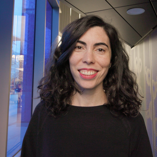
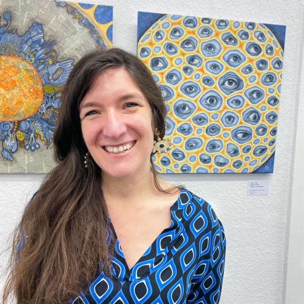
Bio: MartyO, an artist based in California, specializes in creating wearable art, assemblages, and sculptures using salvaged textiles and found items. Transitioning from a background in social work advocacy and law practice, she embarked on a new career as an artist in her fifties. MartyO adheres to zero-waste principles, skillfully transforming discarded, tattered, and stained quilts and linens into distinctive art quilts, garments, and sculptures that convey captivating stories.

With a penchant for taking traditional patchwork and embroidery into the unexpected, MartyO breathes new life into forgotten fabrics. Often the provenance of the textiles remains a mystery, yet her creations give rise to  new narratives. Her art is imbued with emotion as she tackles controversial issues, pays homage to the handwork of others, and inspires others to reduce their carbon footprint.

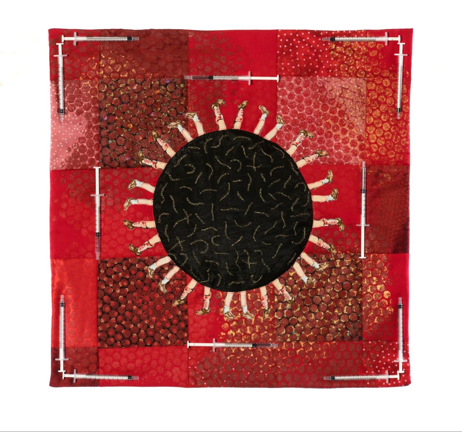
Artist’s Statement:  Southern California artist Marty Ornish creates art quilts, wearable art and assemblages, often incorporating discarded materials to explore themes of sustainability, feminism, history, and science. During her time at UCSD Medical Center in the 1980s, her work as a clinical social worker provided first-hand insight into the art of medicine and the vital role of medical research. Concerned by the rise of vaccine skepticism, she recently created a textile piece advocating for childhood immunizations.

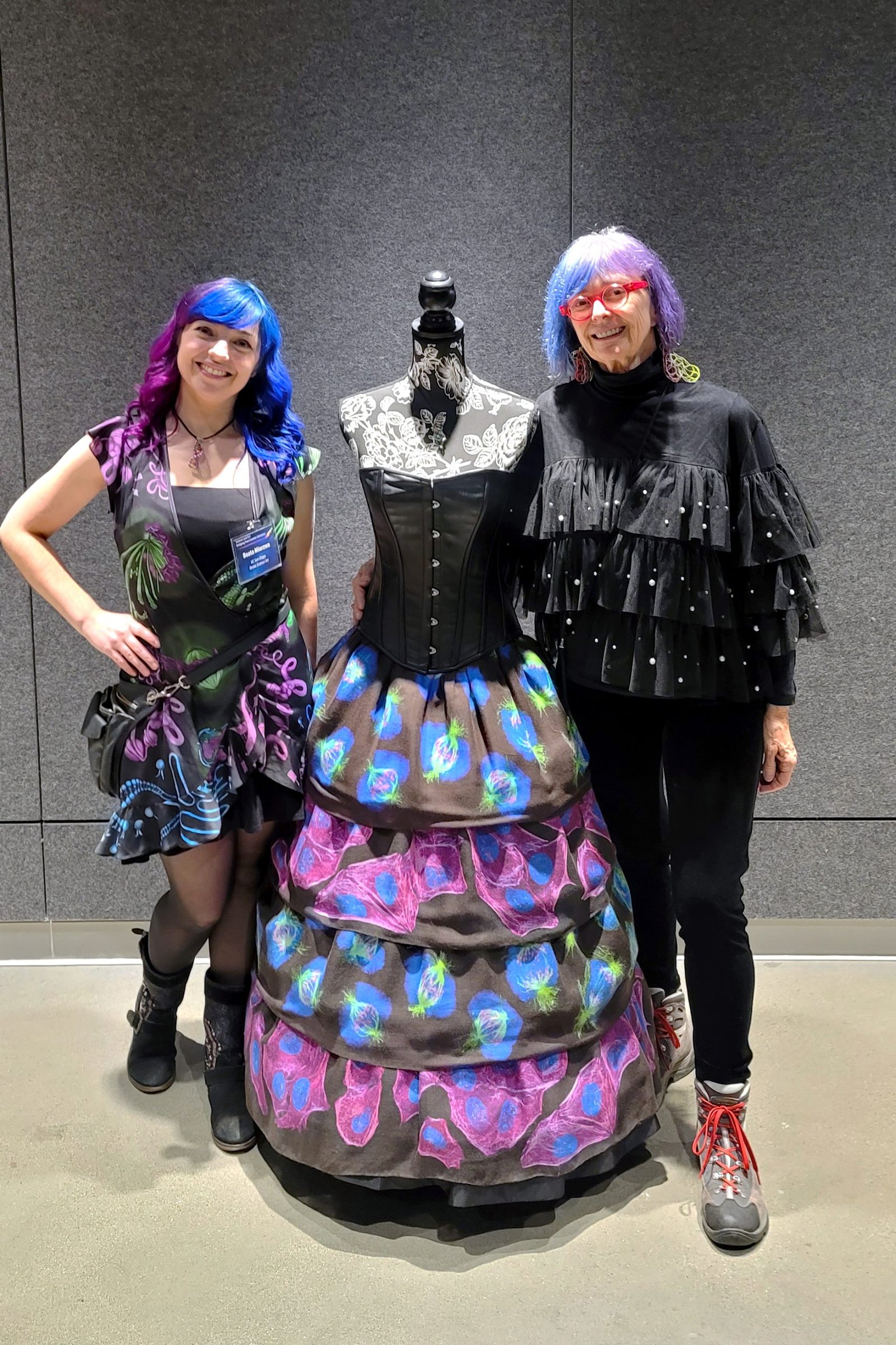
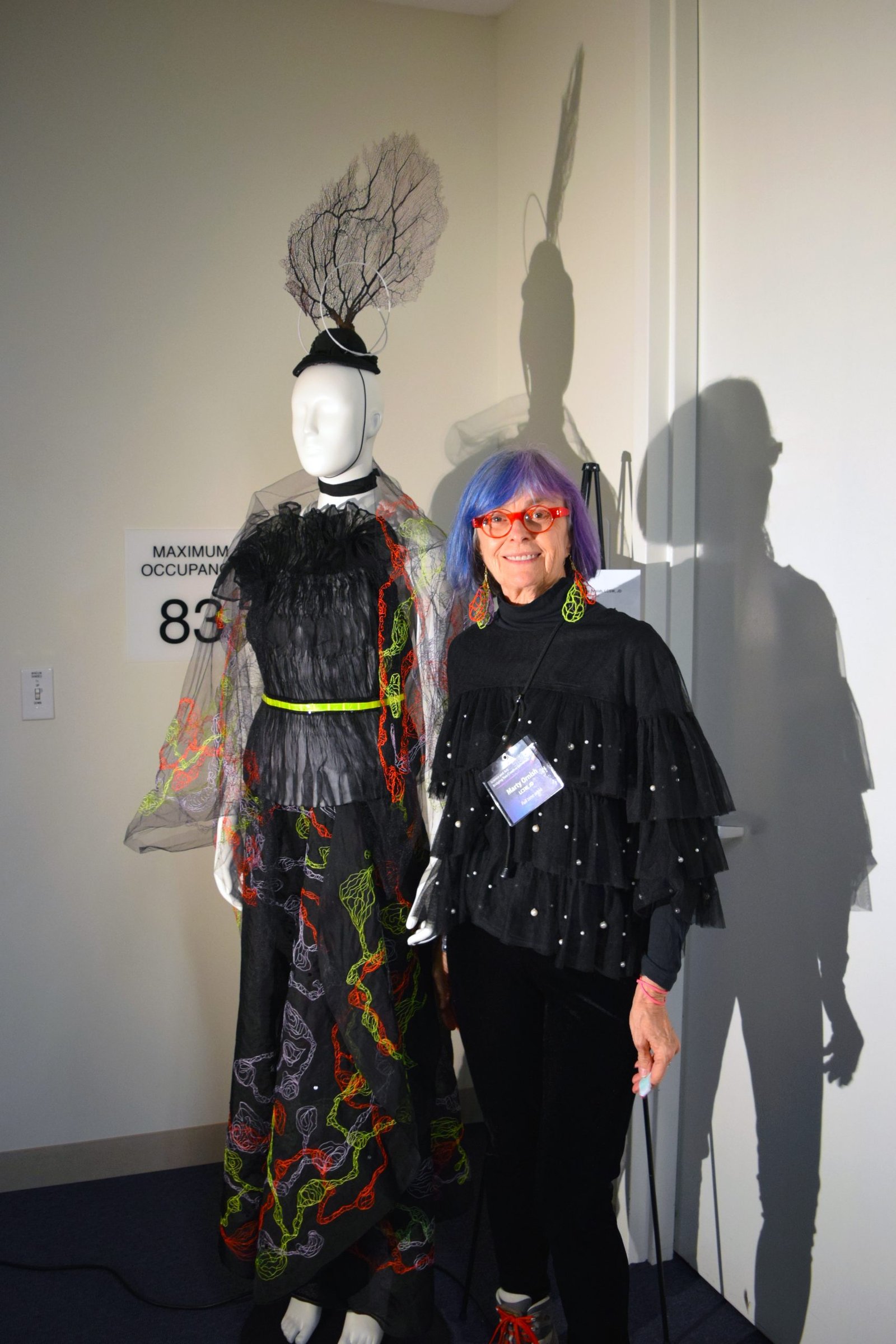
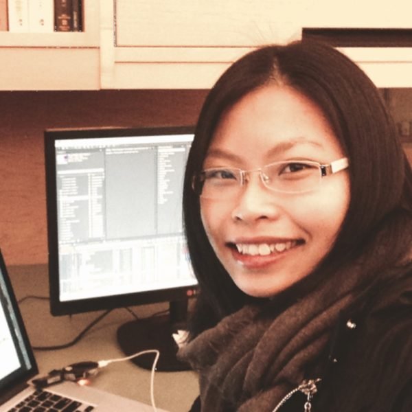
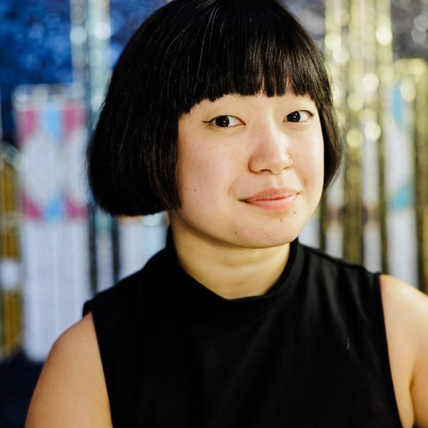
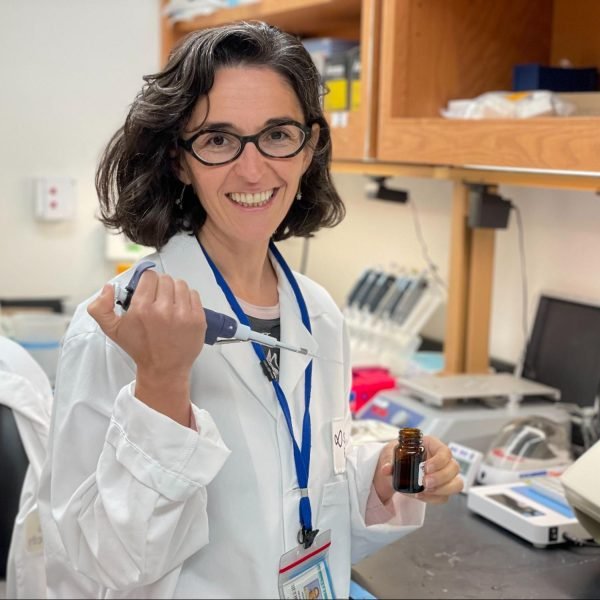
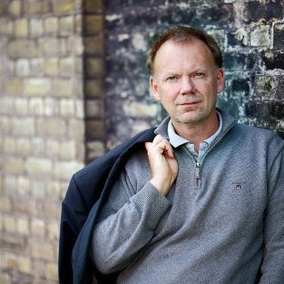
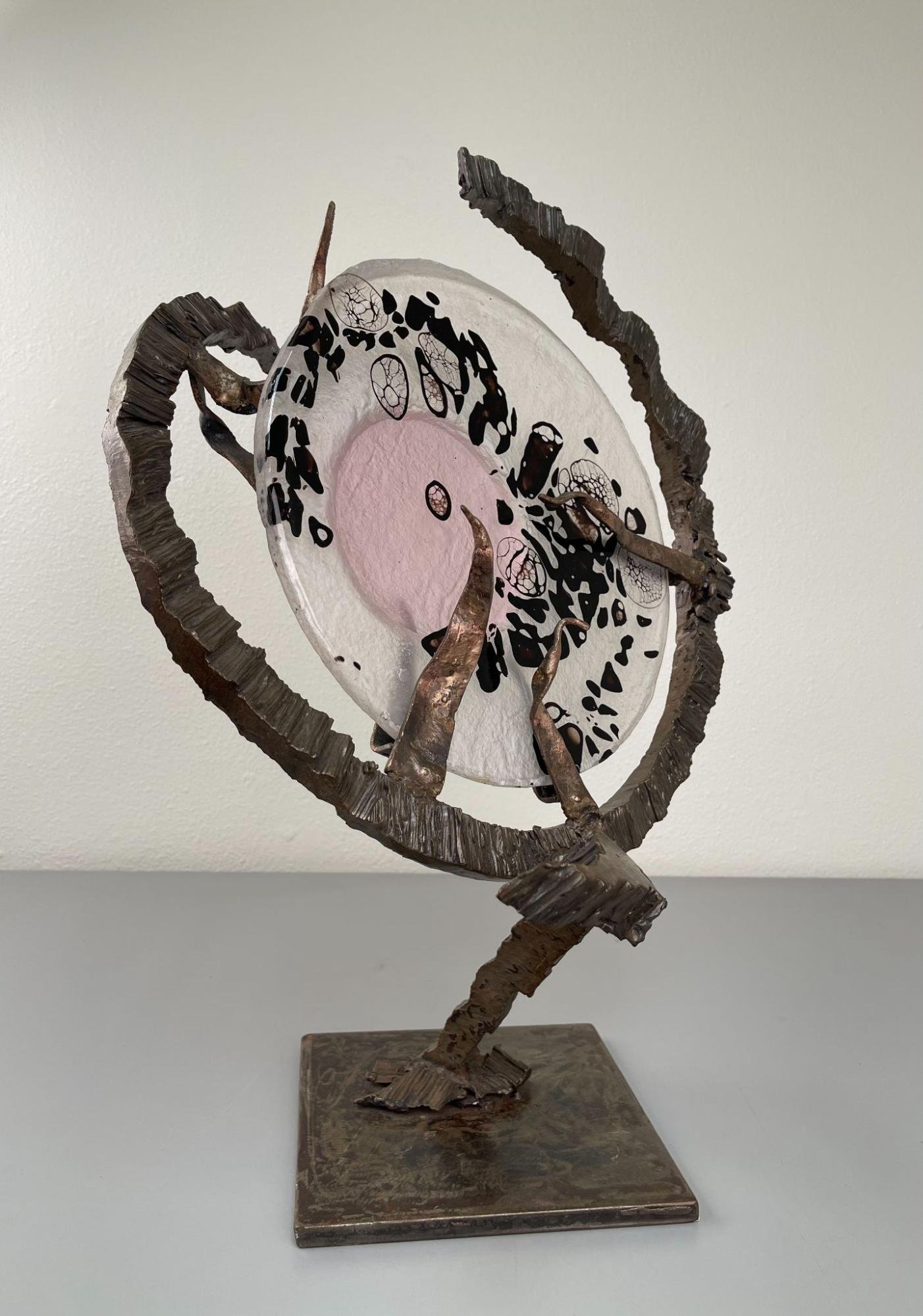
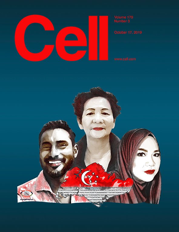
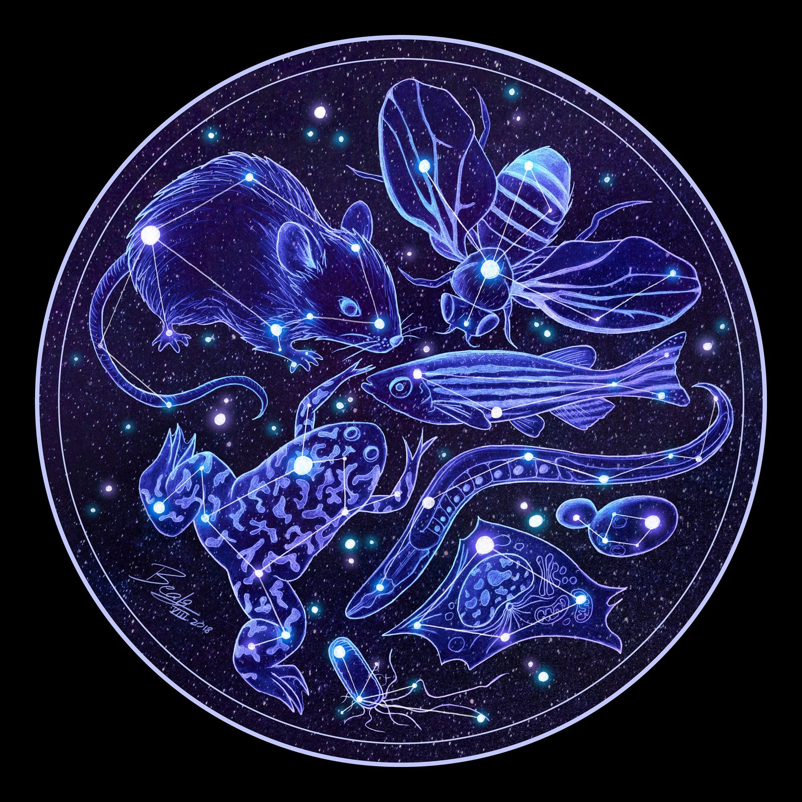
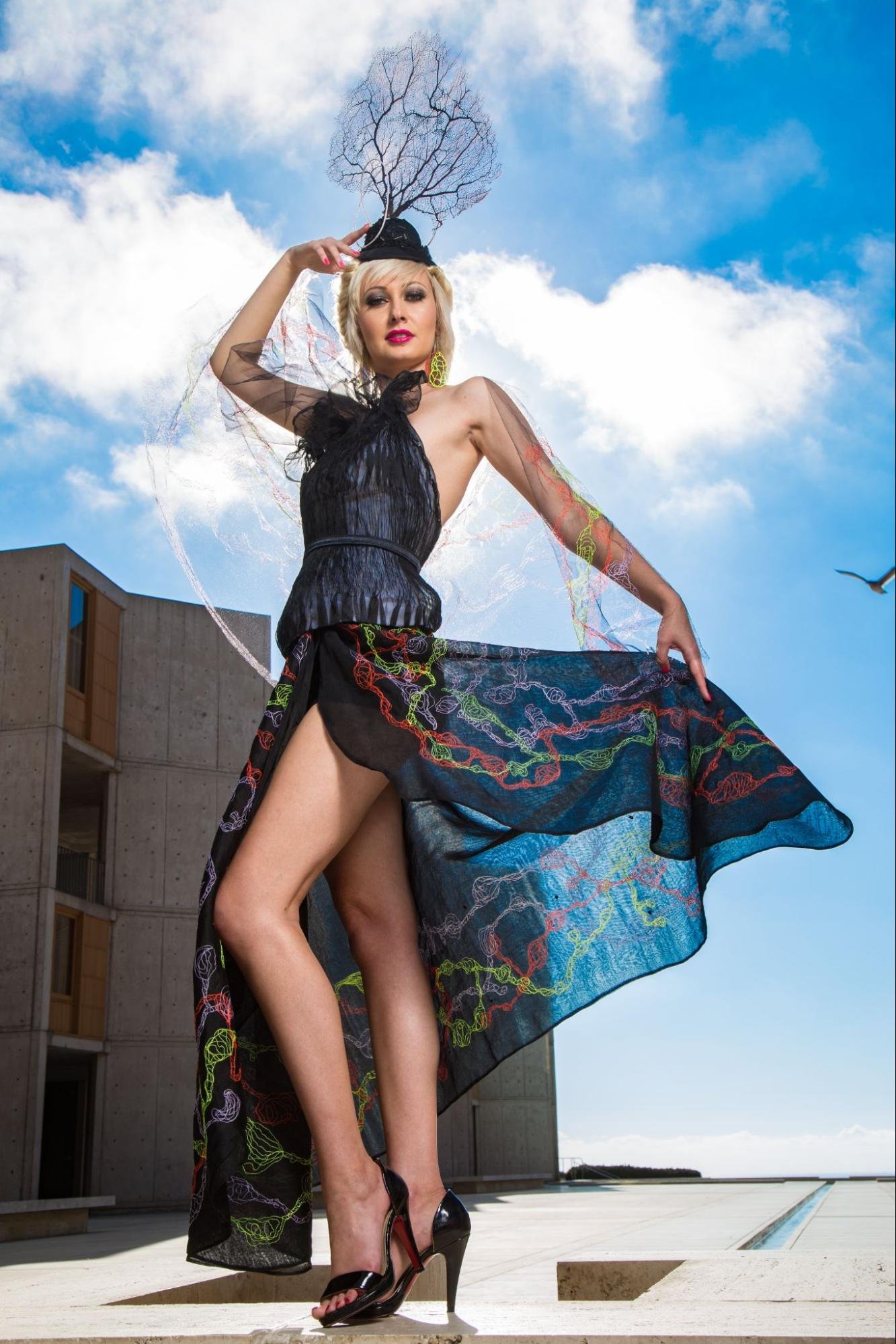
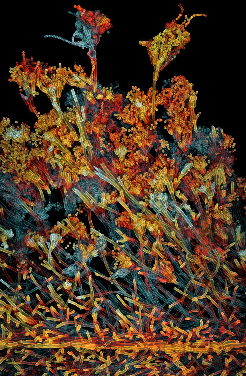
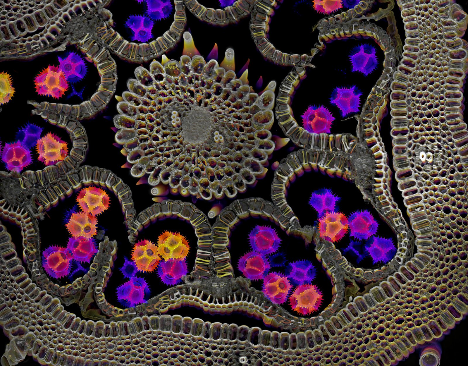
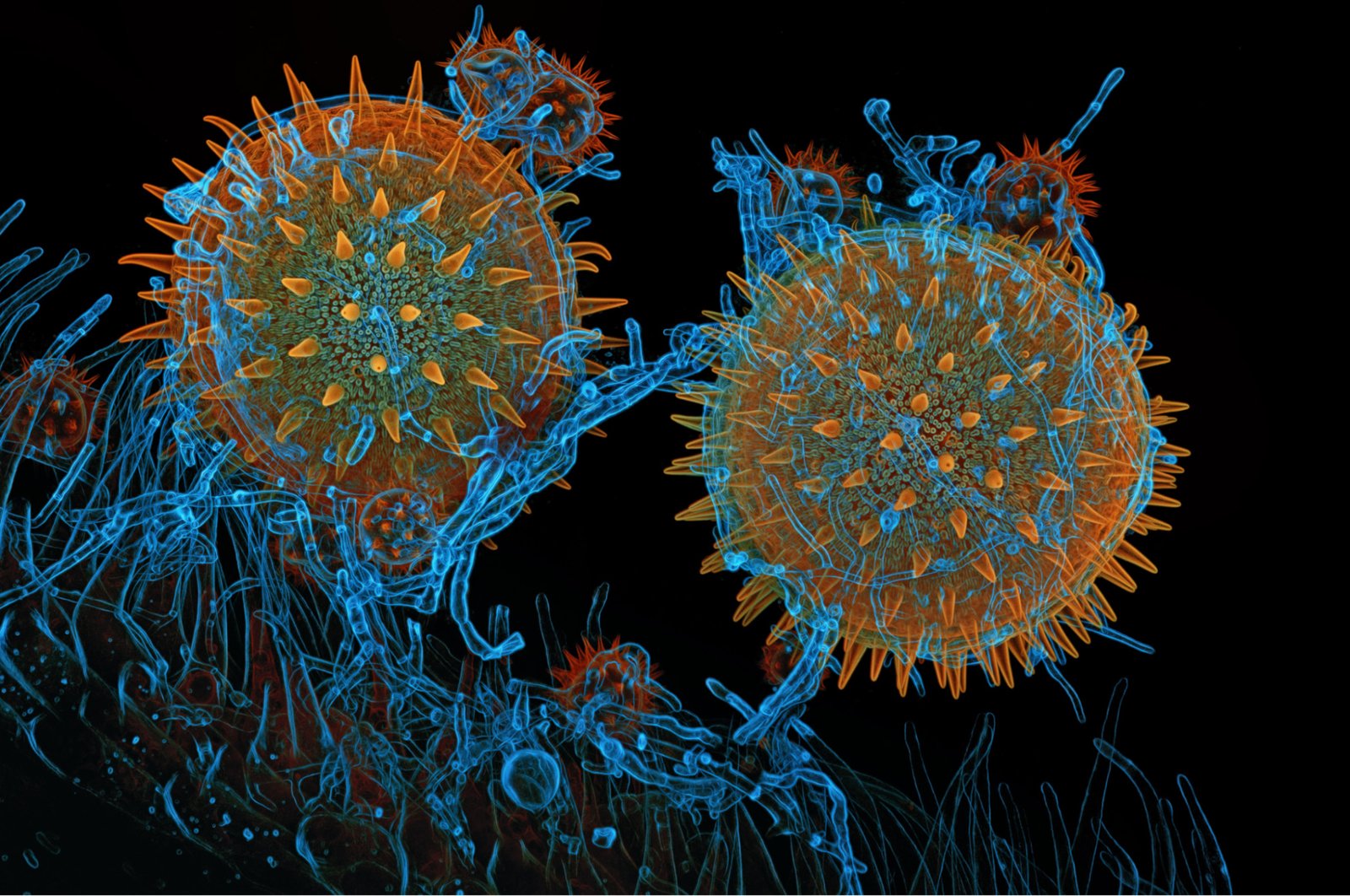
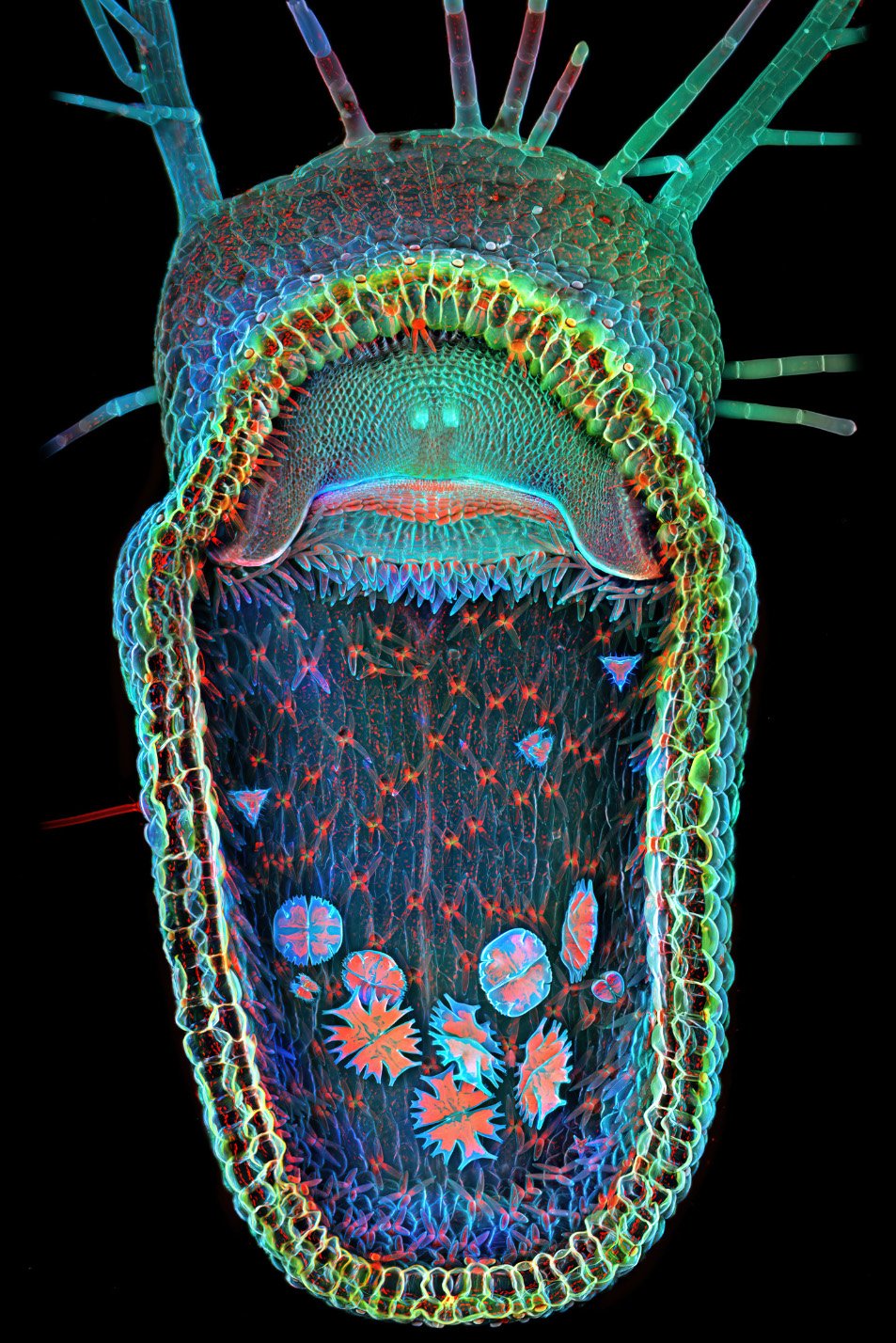
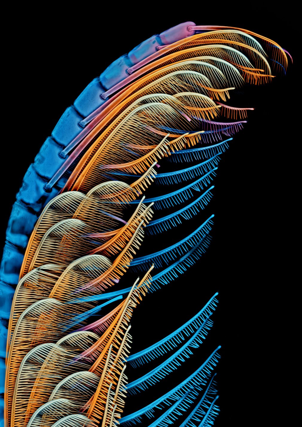
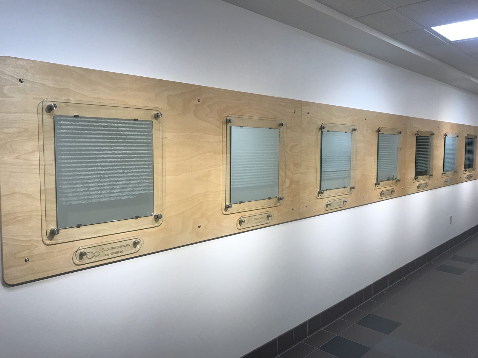
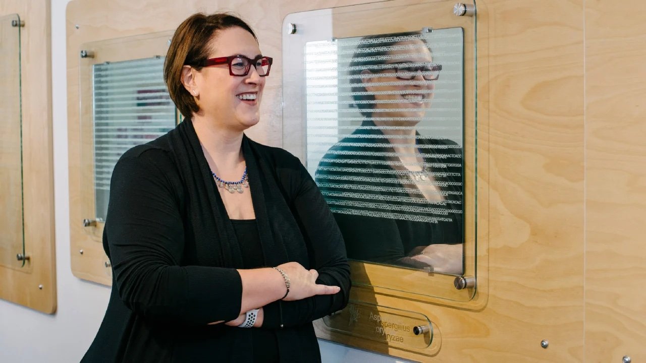
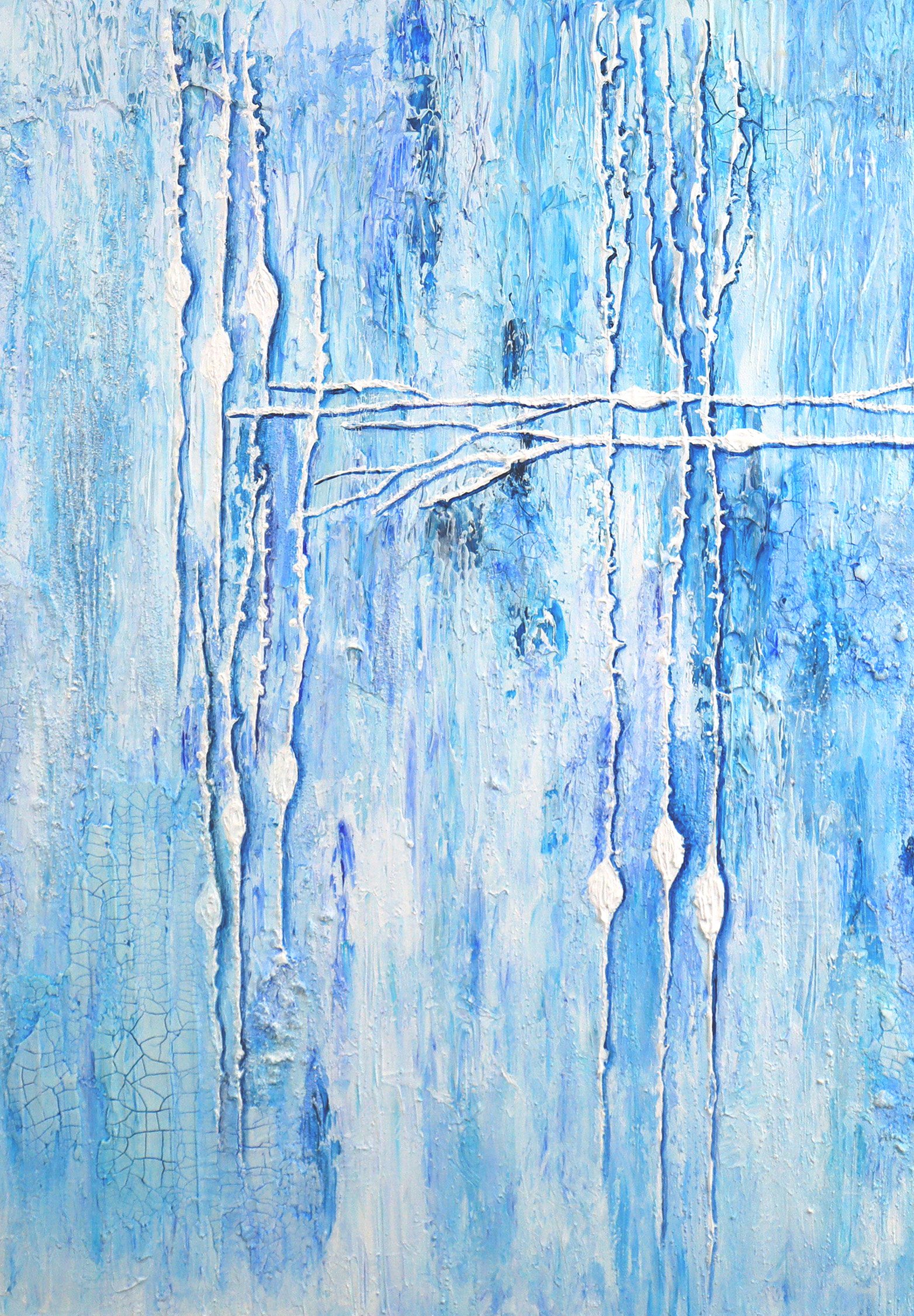
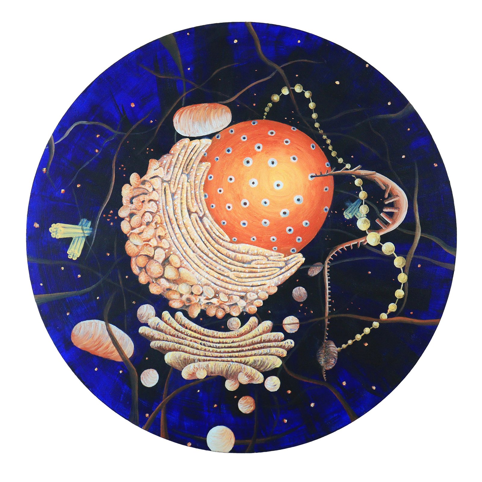
In 2017, the Salk Institute for Biological Studies invited Marty to participate in a fashion show fundraiser supporting women in science. She collaborated with Dr. Cindy Yu Hsin Liu, MD, PhD, a researcher in the Satchidananda Panda Lab, to create Night Visions—a striking departure from her signature use of antique textiles. Using fluorescent thread on silk organza, she rendered cellular structures that dramatically luminesce under black light, fusing scientific precision with artistic expression.

The Power of the Jab (2024)

Artist: Marty Ornish

Commercial cotton fabric remnants, paint, syringes, embroidery thread, syringes, batting

An art quilt made by piecing a selection of red fabrics, stencil painting, hand embroidery, and “fussy cutting” commercial fabric that  promotes the importance of childhood immunizations, with a trapunto cellular structure in the center. Donated syringes create the edges of the piece.

Please contact MartyO for information on usage and permissions.

Night Visions (2017)

Artist: Marty Ornish

Silk Organza, tulle, thread, seed beads, buckram, sea fan, fluorescent spray paint (on the sea fan), felt, cotton, hat combs, floor cement, foam half circle, beading, silk

This dress draws inspiration from a micrograph of the neural network of melanopsin retinal ganglion cells in the eye. The neural network is intricately free-motion embroidered on the skirt and capes using fluorescent thread. Under black light, the embroidery fluoresces dramatically, appearing to float in space. The tightly pleated blouse features an outer layer of black silk organza over white silk organza, subtly glowing to symbolize the interplay of night and day in our circadian rhythms—a homage to the layered structure of the retina. Completing the ensemble is a handmade hat crowned with a large, luminescent coral sea fan, representing the vasculature of the retina.

Please contact MartyO for information on usage and permissions.

B. Duygu Özpolat

Department of Biology, Washington University in Saint Louis

Bio: B. Duygu Özpolat is an assistant professor of biology and a visual artist currently living in Saint Louis, Missouri. Özpolat earned her Ph.D. in Cell and Molecular Biology from Tulane University in New Orleans, and her B.Sc. in Biology from Middle East Technical University in Ankara, Türkiye. She studies stem cells and regeneration, and makes science-themed art (SciArt) inspired by scientific research. For Özpolat, art and science are intrinsically linked through the process of inquiry, experimentation, and technique, for pushing the boundaries of the known and the explored. Özpolat’s artwork and scientific publications can be found at http://ozpolatlab.org

Artist statement: When I look through a microscope, I feel like I am traveling to different worlds that very few people can see. This is a feeling that never gets old. I started drawing and then making ceramics in an attempt to share with others the wondrous things that I observe as a scientist. In my artwork, I use the beauty we encounter in scientific research to celebrate life, cultivate curiosity, and incite appetite for discovery. The seeds of each piece often come from an encounter with an astonishing life form, organ, structure, pattern, or cell.  For my drawings, I dig deeper into the science by reading scientific papers on the subject and examining visual elements more closely before I finally combine them into a drawing. I also like to glean inspiration from traditional art styles (such as Turkish ceramic paintings), natural history museums, and scientific illustrations (especially old biology manuscripts).  Recently, I have been focusing on making pottery forms of organisms that are typically considered scary, yucky, disgusting etc. Segmented worms, insects, spiders, slugs… It is very important to me to invite people to leave their preconceived notions about these animals aside and open their minds to the beauty and wonders of them. We see only a handful of animals (usually vertebrates) celebrated in popular culture when there are literally millions of amazing species out there.

Leech Ice Cream Dish With Spoons (2024)

Artist: B. Duygu Özpolat and Ryan Null

Ceramics

This is a collaboration with my husband Ryan Null, who adores leeches. He designed this plate with the spoons (the baby leeches), and I built it from clay. We picked the glazes together. This is Helobdella austiniensis. They are hermaphrodites, and when they have babies, the babies live attached to the parent’s belly by their suckers. Whenever the parent goes feeding, the babies can reach for the food and feed as well. This species doesn’t suck blood. It eats other animals like insect larvae etc.

Please contact B. Duygu Özpolat for information on usage and permissions.

Worm Candy Dish (2019)

Artist: B. Duygu Özpolat

Ceramics

A nereidid polychaete worm with some artistic license. Ceramic dish at Duygu’s office (it usually has candies inside). Photo credit: Whitney Curtis. (video: https://www.instagram.com/p/Cb5SaX6lKhY/)

Please contact B. Duygu Özpolat for information on usage and permissions.

Blue Membracid (2020)

Artist: B. Duygu Özpolat

Ceramics

A ceramic dish inspired by a colorful membracid (tree hopper). 

Please contact B. Duygu Özpolat for information on usage and permissions.

Peacock Spider (2020)

Artist: B. Duygu Özpolat

Ceramics

Peacock spiders are a type of jumping spiders. They do a silly mating dance shaking their legs, which is depicted in this ceramics piece.  

Please contact B. Duygu Özpolat for information on usage and permissions.

Plankton on Your Plate (2019)

Artist: B. Duygu Özpolat

Ceramics

An oval plate that is decorated with organisms that can be found in a plankton tow from the ocean.

Please contact B. Duygu Özpolat for information on usage and permissions.

Kathryn Peterson

Front Porch Gallery; Fallbrook Art Center; Oceanside Museum of Art; Escondido Arts Partnership; NCSFA; San Diego Watercolor Society; Urban Sketchers San Diego; North County Sketchers

Kathryn Peterson has been creating art all her life, starting in her home state of Alaska. She has lived in many places before settling in San Diego twenty years ago. 

As a promising theoretical mathematics student, she finished her studies with a bachelor’s in fine art. She saw this not as a great departure, but rather a shifted approach to the inquiry of our physical world. 

From remote parts to large cities, she has experienced various countries & cultures. This variety is reflected in her artwork. Kathryn works in various genres and media, finding infinite themes to paint.

Discovery (2024)

Artist: Kathryn Peterson

Acrylic on canvas

From Rosalind Franklin’s break-through x-ray diffraction photo of the double helix to the modern use of touch DNA in forensic science is just a snapshot in time in the story of discovery. This artist’s interpretation of DNA, rather than an accurate model of the double helix, expresses the sense that DNA sheds as simply as Gregor Mendel’s pollen.

This artwork is protected by copyright and cannot be reproduced without the express consent of the artist. Contact her via her website at https://www.kathrynpeterson.org/.

Photo 51 (2012)

Artist: Kathryn Peterson

Collage and acrylic painting on cradleboard

Belatedly we honor the Dark Lady of DNA… Rosalind Franklin’s 1952 51st X-ray diffraction photo of the double helix. James Watson’s self-described reaction re Photo 51: “The instant I saw the picture, my mouth fell open and my pulse began to race.”

This artwork is protected by copyright and cannot be reproduced without the express consent of the artist. Contact her via her website at https://www.kathrynpeterson.org/.

Diatoms (2023)

Artist: Kathryn Peterson

Vintage microscope, watercolor pencil and acrylic, polymer clay, light source, slide of diatoms

Diatom drawings, arranged in visually pleasing patterns, 3D diatom models, sculpted from polymer clay, and actual oceanic diatoms on a slide allow the viewer to engage with the artwork to see these silica-bodied, single-cell organisms that account for 20% of the earth’s oxygen content.

This artwork is protected by copyright and cannot be reproduced without the express consent of the artist. Contact her via her website at https://www.kathrynpeterson.org/.

Stefanie Redemann

University of Virginia, School of Medicine, Center for Membrane; Cell Physiology Department of Molecular Physiology and Biological Physics, Department of Cell Biology, Charlottesville VA

Dr. Stefanie Redemann is an Associate Professor in the Department of Molecular Physiology and Biological Physics at the University of Virginia. She has always been fascinated by how a single cell can become an entire organism, a curiosity that has driven her academic and research career. Dr. Redemann earned her diploma in Biology from the Technical University of Darmstadt in Germany and her PhD in Cell Biology from the Max Planck Institute for Cell Biology and Genetics. Her postdoctoral work at the Medical Theoretical Center in Dresden focused on cell biology and biophysics.

Dr. Redemann’s research centers on understanding the molecular and mechanical principles of spindle assembly and chromosome segregation during cell division. Using advanced imaging technologies and genetic tools, her work aims to uncover the mechanisms behind these critical cellular processes, which have significant implications for cancer biology and reproductive health. 

Dr. Redemann’s journey to becoming a scientist was not straightforward. After high school, she initially worked in a pharmaceutical company before deciding to pursue higher education. Encouraged by her boss, she went on to study Biology at the Technical University of Darmstadt and later joined the lab of Dr. Hyman at the Max Planck Institute for her PhD. Her passion for science and her dedication to interdisciplinary collaboration have been key drivers in her career. In 2018, she moved to the United States to start her own independent research group at UVA, where she continues her research and to mentor the next generation of scientists

The provided Artwork is the result of her research, using 3D electron tomography to study the assembly of mitotic and meiotic spindles, the machinery that segregates Chromosomes during cell division.

Going Through C.elegans Meiosis (2018)

Artist: Stefanie Redemann

Meiosis is a special type of cell division that produces reproductive cells, such as sperm and eggs, in sexually reproducing organisms. It involves two rounds of division that result in four cells, each with half the number of chromosomes as the original cell. This process is crucial for genetic diversity and proper chromosome number in offspring.

This video showcases 3D reconstructions of the meiotic spindle in the roundworm C. elegans. The meiotic spindle is a structure made of microtubules and chromosomes that plays a key role in cell division. Different stages of meiotic division, from metaphase to various phases of anaphase (early, mid, and late) are shown.

The reconstructions are created from electron tomograms of 300nm thin sections of high-pressure frozen and plastic-embedded C. elegans oocytes (egg cells). By segmenting and stitching together the microtubules from these sections, we form a large-scale 3D image that reveals every single microtubule in the meiotic spindle.

Please contact Stefanie Redemann (sz5j at virginia.edu) for information on usage and permissions.

Inside a Drosophila Oocyte (2024)

Artist: Stefanie Redemann

Intracellular transport is the process by which molecules and organelles move within a cell. This movement is essential for various cellular functions, including nutrient distribution, waste removal, and communication between different parts of the cell. Microtubules, which act like train tracks, play a crucial role in this transport system, guiding vesicles and organelles to their destinations.

This video showcases 3D reconstructions of a 10µm x 10µm area in the oocyte (egg cell) of the fruit fly Drosophila melanogaster. Upon fertilization, these oocytes will develop into eggs and eventually new fruit flies.

The reconstruction is created from electron tomograms of 200nm thin sections of a high-pressure frozen and plastic-embedded Drosophila oocyte. By segmenting and stitching together the microtubules from these sections, we form a large-scale 3D image that reveals every single microtubule (in green and purple) and organelle. Mitochondria, the powerhouses of the cell, are shown in red, while other vesicles necessary for transport and degradation of cellular components are shown in yellow. As transport in cells happens along microtubules, similar to train tracks, the microtubules that come in contact with the vesicles are highlighted in purple.

Please contact Stefanie Redemann (sz5j at virginia.edu) for information on usage and permissions.

Rotating Mouse Meiosis Chromosomes (2024)

Artist: Stefanie Redemann

Metaphase is a stage of cell division where chromosomes align in the center of the cell, forming what is known as the metaphase plate. This alignment ensures that each new cell will receive an identical set of chromosomes. Chromosomes are structures made of DNA and proteins that carry genetic information crucial for the development and functioning of living organisms.

This video presents a 3D tomography of chromosomes aligned on a metaphase plate, rotating to showcase their alignment. The data is derived from a metaphase II mouse meiotic spindle, highlighting the precise organization of chromosomes during this critical stage of cell division.The reconstruction is created from electron tomograms of 200nm thin sections of high-pressure frozen and plastic-embedded Mouse oocytes. 

In the video, chromosomes are depicted in various colors to distinguish them from one another, providing a clear view of their arrangement and structure. 

Please contact Stefanie Redemann (sz5j at virginia.edu) for information on usage and permissions.

C. elegans Mitotic Spindle (2017)

Artist: Stefanie Redemann

Mitosis is a type of cell division that results in two daughter cells, each with the same number of chromosomes as the parent cell. This process is essential for growth, development, and tissue repair in multicellular organisms. During mitosis, chromosomes are duplicated and evenly distributed to ensure that each daughter cell receives an identical set of genetic information.

This video showcases a 3D reconstruction of the metaphase stage mitotic spindle in the roundworm C. elegans. Metaphase is a critical phase where chromosomes align in the center of the cell, forming the metaphase plate. This alignment ensures that each daughter cell will receive an identical set of chromosomes.

The reconstructions are created from electron tomograms of 300nm thin sections of a high-pressure frozen and plastic-embedded C. elegans embryo. By segmenting and stitching together the microtubules from these sections, we form a large-scale 3D image that reveals every single microtubule in the mitotic spindle. Kinetochore microtubules, which attach to the chromosomes and help pull them apart, are shown in orange. These specialized microtubules play a crucial role in ensuring accurate chromosome segregation during cell division.

Please contact Stefanie Redemann (sz5j at virginia.edu) for information on usage and permissions.

Alex Ritter

Altos Labs, Inc.

Alex Ritter is a scientist and glassworker based in San Francisco, California. After growing up and attending college in North Dakota, Alex went on to pursue his PhD training at University of Cambridge, where he developed specialized microscopy techniques to capture videos of killer immune cells as they engage and destroy cancer. Alex’s scientific work has been published in top journals including Science and Nature. His videos are widely used to illustrate the activity of killer T cells in textbooks, television, scientific talks, and online. Outside the lab, Alex creates art using the medium of glass to sculpt 3-dimensional models of the cells that he observes in the microscope. He currently oversees work as a senior scientist at a fast-growing biotechnology company called Altos Labs.

T Cells Killing Cancer Under the Microscope (2021)

Artist: Alex Ritter

Spinning disk laser fluorescence confocal microscope

We have an army of killer immune cells inside of us that patrol our tissues, searching for virus-infected and cancerous cells. When these immune cells, called cytotoxic T cells, detect a deviant target cell, the T cell engages and injects potent toxins into the target. This is a short video captured in a microscope of actual T cells (magenta) swarming a cancer cell (cyan) to engage and destroy it. The red wave indicates the precise moment that toxins are delivered to the target cell. This unique cell-cell interaction has been called the “kiss of death”.

Please contact [alexritter09 at gmail.com] for information on usage and permissions.

Kiss of Death (2023)

Artist: Alex Ritter

Sculpted glass

The artist has spent his career studying this interaction of cytotoxic T cell and target cell using live cell microscopy, and represents this interaction in hot sculpted glass. In this way, the artist is representing what he sees in the microscope using the medium of glass. The hope is that we can connect with the biology inside of us and recognize the good work our immune cells do every day to protect us from viral and cellular threats.

Please contact [alexritter09 at gmail.com] for information on usage and permissions.

T Cell, Engaged (2022)

Artist: Alex Ritter

Sculpted glass

The artist has spent his career studying this interaction of cytotoxic T cell and target cell using live cell microscopy, and represents this interaction in hot sculpted glass. In this way, the artist is representing what he sees in the microscope using the medium of glass. The hope is that we can connect with the biology inside of us and recognize the good work our immune cells do every day to protect us from viral and cellular threats.

Please contact [alexritter09 at gmail.com] for information on usage and permissions.

Michael Rosnach

Michael Rosnach is an artist and industrial designer working at the Disease Biophysics Group (DBG), Harvard, a heart-focused research group headed by Professor Kevin Kit Parker. 

Michael comes from academic figurative drawing and painting, having studied at the New York Academy of Art, but his current focus is on bringing art into the science world, and art into the science world.  As part of that goal he makes artworks based on his experience at the lab, the narrative of the scientists’ experimental work, the unfamiliar and novel objects created in the lab, and the profound difference between the narrative and workflow of artists and scientists.  Much of his work revolves around talking to scientists, discussing with them the narrative of their projects, and creating graphic and artistic works to visually communicate the science of the DBG to the audience.

Having largely left the realm of pure studio painting, Michael also  works directly with the tools and technology found in the lab, and looks for ways to link the barely explored edge of the natural world with the human world of art. 

David Schneider

Stanford University, Department of Microbiology and Immunology

I study the pathology of infectious diseases, and I’m trying to understand why we get sick when we become infected.  After upsetting some people when I proposed some biology that they thought was impossible, I figured the problem was that I didn’t describe it well enough (but that wasn’t the problem).  I started illustrating my work using stop motion animation that looked at infections in a humorous way.  The downside of that is I had to show it on a screen.  I decided to start making small sculptures of my work that I could carry with me as visual aids.  I’m currently trying to explain the shape of “disease space” that different hosts can occupy.  My data and my models show that this space is tetrahedral and so I’ve been making a variety of linear, triangular and tetrahedral objects.  I love the color and I’ve been using glass in the form of enamel or cast material in my sculptures. 

Data Waves (2017)

Artist: David Schneider

Plywood and walnut

This automaton shows the progression of 8 symptoms as a mouse is infected and then recovers from malaria.  The cams are circular projections of the timeseries of cytokines, metabolites, circulating cells and red blood cell concentrations.  This is the best way I’ve found of showing that an infection produces a wave-like disturbance that passes through our physiology and then disappears.  It gives me hope that instead of describing all of these different parameters that someday I will be able to describe the amplitude, wavelength and velocity of the physiological wave.  

Please contact David Schneider for information on usage and permissions.

Organs of Interest (2024)

Artist: David Schneider

Plique a jour – enamel, silver, brass, wood, glass

This is a concept for organ jewelry.  I would like a set of jeweled organs that I can wear for when I have to describe the physiological changes that I study.  I made most of the important ones I study.  I’m not sure how to wear all of these at once.  

Please contact David Schneider for information on usage and permissions.

A Mischief of Mice (2024)

Artist: David Schneider

Glass, dichroic glass, silver

These glass mice represent the diverse mice that I study.  They differ from each other in many ways, but when you look at them just right you can see an underlying triangular pattern to their distribution.  This is a work in progress.  The mice are going to be used for two projects.  The first will be a tetrahedral mobile and the second a stained glass window.

Please contact David Schneider for information on usage and permissions.

Optimal Mice (2023)

Artist: David Schneider

Plique a jour – glass, LED

I use these mice to help explain Pareto optimization.  Before I had these I would wave my hands in the air to try to describe that idea.  Now I can hold up a glowing mouse that is easily visible from the back of an auditorium and give a clear explanation.  The idea is that if you have two well adapted states then selection for both these states will produce individuals on a line that connects the two.  If you have three states, you will select a triangle of phenotypes.

Please contact David Schneider for information on usage and permissions.

Pocket Cell (2022)

Artist: David Schneider

Plique a jour – enamel, silver

This is a pocket sized cell I can pull out whenever I need to talk about some cell biology.  I wanted it to look like the modern illustrations of cells that use internal reflections, but to make this as a physical object rather than something that existed on a screen. 

Please contact David Schneider for information on usage and permissions.

Igor Siwanowicz

Janelia Research Campus of the Howard Hughes Medical Institute

He earned his PhD at the Max Planck Institute in Germany in structural biochemistry, but his love for animals and nature, as well as a desire to see the bigger picture, drove him to refocus his scientific work on neurobiology. This transition was facilitated by his expertise in invertebrate morphology, which he developed through macrophotography. A confocal microscope became his key tool of trade, allowing for even more intimate insight into natural forms than is possible through a macro lens. For the past 12 years, he has been studying the anatomy and biomechanics of movement in invertebrates at Janelia. His images have placed a total of 30 times in Nikon’s Small World, Olympus’s BioScapes, and Evident Image of the Year competitions. In 2020, he received the Royal Photographic Society’s Scientific Imaging Award. Siwanowicz’s science-informed art aims to awaken viewers to the beauty of natural forms and multiple facets of design present in nature at every scale.

Aspergillus Mold (2024)

Artist: Igor Siwanowicz

Matte fine art paper

Confocal image of an Aspergillus sp. Cultured on an agar plate. Magnification: 400x. Depth color-coded projection.

Freely available for use under a CC-BY-4.0 license.

Common Chicory Foret with Developing Pollen Grains (2023)

Artist: Igor Siwanowicz

Matte fine art paper

Cross section through a common chicory floret showing pollen grains developing inside of the anthers. Technique: confocal. Magnification: 400x. Depth color-coded projection.

Freely available for use under a CC-BY-4.0 license.

Mallow Pollen Infected by a Fungus (2024)

Artist: Igor Siwanowicz

Matte fine art paper

The image shows two mallow pollen grains being consumed by a filamentous fungus while attempting to germinate on the flower’s stigma. The smaller spiky balls are pollen grains of an aster. Technique: confocal. Magnification: 400x

Freely available for use under a CC-BY-4.0 license.

Humped Bladderwort Trap Interior (2012)

Artist: Igor Siwanowicz

Matte fine art paper

The image shows the inside of a trapping organ, or a bladder, of an aquatic carnivorous plant, Urticularia gibba. The intricate shapes inside it are single-cell freshwater algae, desmids. Technique: confocal. Magnification: 100x.

Freely available for use under a CC-BY-4.0 license.

Antenna of a Mole Crab (2023)

Artist: Igor Siwanowicz

Matte fine art paper

This colorful, feathery stalk is the antennae of a mole crab, also known as a sand flea. You may have seen these little crustaceans burrowing into the sand at the beach as the waves wash over the shore. They use these antennae to filter food from the water and sweep it into their mouths. Technique: confocal. Magnification: 100x. Depth color-coded projection.

Freely available for use under a CC-BY-4.0 license.

Planthopper Parasite Moth Caterpillar (2012)

Artist: Igor Siwanowicz

Matte fine art paper

This is a ventral view of the caterpillar stage of a planthopper parasite moth (Epipyrops exigua). These caterpillars are unique in the Lepidoptera order that includes moths and butterflies in that they are ectoparasites; that means they attach themselves to the body of another insect, typically a planthopper (in the infraorder Fulgoromorpha), and feed on their circulatory fluid, known as hemolymph, as they grow. Technique: confocal. Magnification: 100x.

Freely available for use under a CC-BY-4.0 license.

Damselfly Flight Muscle (2021)

Artist: Igor Siwanowicz

Matte fine art paper

The image shows a cross section through a damselfly (Argia sp.) forewing depressor muscle. The radial arrangement of the muscle’s components is striking: actin-rich lamellar (slab-like) myofibrils (teal) surround the central core of nuclei (yellow). The spaces between myofibrils are occupied by mitochondria, or sarcosomes. Axons of motorneurons innervating the muscle as are shown in red. Technique: confocal. Magnification: 630x.

Freely available for use under a CC-BY-4.0 license.

Tongue of a Snail (2021)

Artist: Igor Siwanowicz

Matte fine art paper

The image shows a radula, or a rasping tongue, of Elimia virginica, a snail endemic to the lower Potomac and her tributaries. Technique: confocal. Magnification: 250x. Depth color-coded projection.

Freely available for use under a CC-BY-4.0 license.

Ahna Skop

UW-Madison Genetics, Affiliate faculty in the Art Department and Life Sciences Communication

Ahna Skop is a geneticist, artist, and professor at the University of Wisconsin-Madison, known for her groundbreaking research on cell division and the midbody and midbody remnant(MBR). Her lab focuses on understanding asymmetric cell division mechanisms, with particular emphasis on the role of the MBR  in cell fate, tissue polarity, and oncogenesis. In 2004, Skop pioneered proteomic and genomic approaches to identify novel cell division proteins, publishing her findings in Science. Recently, her lab discovered that the midbody is a translationally active, RNA-containing organelle, potentially functioning in the spread of cancer.

Skop’s unique background as the child of artists has significantly influenced her scientific approach, inspiring her to combine art and science in innovative ways. She has curated scientific art exhibitions, created a 40-foot scientific art piece called “Genetic Reflections,” and authored a coloring book showcasing the beauty of genetics. Skop is also passionate about increasing diversity in STEM fields, having received numerous awards for her inclusive teaching and outreach efforts. Her scientific and artistic achievements have been recognized with prestigious honors, including the Presidential Early Career Awards for Scientists and Engineers (PECASE) and an AAAS IF/THEN Ambassadorship.

Genetic Reflections Coloring book: https://skoplab.weebly.com/genetic-reflections.html
Ahna Skop Science Art: https://skoplab.weebly.com/science-art.html

Genetic Reflections: A Coloring Book (2020)

Artists: Elif Kurt, Caitlin Marks, Ahna Skop

Book

Genetic Reflections – A Coloring Book aims to inspire young students and the public to explore the beauty of science and genetics. The organisms in this book are considered ‘model’ organisms, as they are widely studied in laboratories with hopes to understand human biology, disease pathologies, and ways to improve agricultural crops. Despite the great differences in shape and size, on the genetic level there are lots of similarities.In every species, DNA sequences consist of the same four building blocks (G, C, A, and T). However, slight changes in their use, even in the same gene, can occur in each species. The way our bodies and cells work are well conserved throughout evolution, even in species that may look very different from us. The beauty of our world, even on the cellular level, is apparent.Genetic Reflections – A Coloring Book is a collaboration between Ahna Skop, Elif Kurt and Caitlin Marks; two UW-Madison undergraduate Skop Lab members. This coloring book is the outcome of a year-long independent study in Life Sciences Communication with goals to broadly disseminate the Genetic Reflections scientific glass art installation created by Angela Johnson and Ahna Skop.Part of the proceeds of this book will be donated to charities and programs that support STEAM (Science, Technology, Engineering, Arts, and Mathematics) educational innovations or public outreach events.

For Purchase: https://a.co/d/1eA8bFK

Please contact Ahna Skop on usage and permissions.

Genetic Reflections Art Piece

Artists: Ahna Skop & Angela Johnson

Sandblasted glass, mirrors, wood

Genetic Reflections is a public art installation and coloring book that aims to educate the public about genetics and science.

Located outside the Genetics/Biotechnology Center auditorium at UW–Madison, this installation features glass panes with the letters of the genetic code laser-etched into them. The panes are mounted on mirrors and illuminated by angled light. The installation was created by artist Angela Johnson and genetics professor Ahna Skop

Link: https://skoplab.weebly.com/genetic-reflections.html

Please contact Ahna Skop on usage and permissions.

Kathleen Sluka

Professor, University of Iowa

I have always been interested in art, and started painting at a young age. Despite this interest, I pursued a career in healthcare and science, a career I truly enjoy. I trained as a physical therapist and then obtained a PhD in Anatomy, which was followed by a faculty position at the University of Iowa. For years, I did not pursue art but rather spent time focusing on building a successful scientific career. Currently, I run an active research program, part of my day job as a Professor at the University of Iowa, focusing on the neurobiology of musculoskeletal pain and analgesia. However, about a decade ago I again began painting and experimenting with art – this slowly evolved into science art. For the last several years I have exclusively focused my art on bringing the beauty of biology to canvas in normal and disease states. I have exhibited my art at local venues, and several of my paintings have been selected as covers of scientific journals. 

Analgesia (Pain Relief)

Artist: Kathleen Sluka

Acrylic on canvas, 24x30”

Analgesia represents the opposite of pain. While pain is associated with chaos and inflammation, fear, anger and stress, pain relief is associated with order, calm, and peace. The piece was designed with blues and white and used a variety of textures to bring simulate “ice”, which has been used for centuries to control pain. 

Please contact Kathleen Sluka for information on usage and permissions.

Microuniverse

Artist: Kathleen Sluka

Acrylic on canvas, 24” round

The inside of the cell is an amazing universe of structures and molecules that provide life. The cell, in its nucleus, holds life’s code (DNA) and through interaction with several structures converts that code to the proteins that that provide function.

Please contact Kathleen Sluka for information on usage and permissions.

Mondrian Musculoskeletal

Artist: Kathleen Sluka

Acrylic on canvas

The musculoskeletal system is composed a variety of tissue types designed to promote movement. This painting is designed based on Piet Mondrian’s style using primary colors and geometric shapes. Within each square I show a tissue or cell type related to the musculoskeletal system in primary colors. These include, tendon, muscle, bone, connective tissue, synovium, nerve, cartilage, and muscle tissue.

Please contact Kathleen Sluka for information on usage and permissions.

Pain Gate

Artist: Kathleen Sluka

Acrylic on canvas

The Gate Control Theory of Pain published in 1965 theorized a balance between excitation and inhibition within the central nervous system. Signals from the sensory neurons (boulders) send signals to the central nervous system (trees) through to modulate pain. The gate can open and close due to activity in different type of neurons.

Please contact Kathleen Sluka for information on usage and permissions.

Pain Brain

Artist: Kathleen Sluka

Multimedia

Brain imaging studies have transformed our understanding of how pain is processed in the brain. This multimedia collage uses brain images and text from published studies by pain scientists.

Please contact Kathleen Sluka for information on usage and permissions.

Speed Demons

Artist: Kathleen Sluka

Acrylic on canvas

Neurons send electrical signals through their axons, in brown. The speed of the electrical signal is determined by the myelin sheath which is made from a glial cell called an oligodendrocytes (blue). The myelin is considered insulation and speeds the conduction of electrical signals down the axons.

Please contact Kathleen Sluka for information on usage and permissions.

Rou-Jia Sung

Carleton College

As a structural biologist, I have always been fascinated by the beauty of molecular structure. Visualizing how atoms connect together to form complex molecular machines has implications for understanding how biological processes work and appreciating the phenomenal visual complexity of these molecules. Seeing the intersection between science and art has been an important part of my teaching– whether it is creating molecular visualizations or playing with bioluminescence, thinking of creative ways for them to combine art and science has always been a passion of mine!

Secondary Structure (2017)

Artist: Rou-Jia Sung

Paint on tile, glazed and fired

Painting of secondary structure on ceramic tile.

Please contact Rou-Jia (rsung[at]carleton.edu) for information on usage and permissions.

Clathrin Coated Pit (2018)

Artist: Rou-Jia Sung

Plastic

3D printed rendering of a clathrin coated pit.

Please contact Rou-Jia (rsung[at]carleton.edu) for information on usage and permissions.

Ayelen Valko

GEDOK, Heidelberg

Ayelen is an Argentinean cell biologist and artist. She received her Ph.D. from the Leloir Institute in Buenos Aires, specializing in microscopy techniques that allowed her to access the fascinating subcellular universe. With the goal of artistically conveying the beauty hidden in cells, and in parallel to her scientific training, she was trained in various artistic techniques at the National University of Arts in Argentina. She then specialized in scientific and naturalistic illustration at institutions such as the “Ernst Haeckel” Laboratory of Scientific Painting, the “Adumbratio” Center of Scientific Illustration, and others. Several of her works have been selected as cover for various scientific journals, including the Journal of Cell Science, Autophagy Journal, Autophagy Reports, Bio-protocol, and Developmental Biology. She extensively described two of these scientific paintings in a commentary article published in the Autophagy Journal (Vol 15, Issue 11).

Her artworks inspired by the cellular universe have been exhibited in galleries, museums and scientific institutions in Argentina and Europe, including the Quinquela Martin Museum and the Rómulo Raggio Foundation in Buenos Aires, the Complexity Science Hub in Vienna, the Max Planck Institute for Biophysics in Frankfurt, and the GEDOK Gallery in Heidelberg, Germany. Currently, Ayelen is based in Heidelberg, where she has become a member of GEDOK, a community of women artists and art patrons. This artistic organization – with locations in 23 German cities – is the oldest and largest network for women artists in Europe. Ayelen focuses on organizing and curating art shows, creating science-inspired covers for scientific journals, and leading science communication activities through art.

Anthropomorphic Interpretation of Lysosome 1 (2023)

Artist: Ayelen Valko

Oil on canvas

This painting illustrates the intrinsic processes of biological destruction and catabolism associated with the lysosome – the ‘stomach’ of the cell. Ayelen often incorporates textures from dreams and memories into her paintings, adding a personal depth to each science-inspired piece. In this oil on canvas, she has taken it a step further by introducing suffering human beings into the composition. Her hypothesis is that viewers will grasp the biological message more easily than in similar pieces without human representation. Organic forms are a signature of Ayelen’s art. In this case, she has created a complex organic matrix that surrounds the anthropomorphic figures trapped inside the lysosome. She created these textures over a long period of time as an attempt to clear her mind and translate onto canvas whatever her unconscious mind dictated. So these forms encapsulate the joys and frustrations of daily life. To create this painting, she drew inspiration from Hieronymus Bosch’s depictions of Hell and Heaven, H.R. Giger’s biomechanical universes, Gaetano Zumbo’s dramatic sculptures, Frida Kahlo’s symbolic paintings, Remedios Varo’s surreal creatures, and numerous other luminaries of the Surrealist movement. This work has a palette and composition reminiscent of transmission electron microscopy, a technique that has allowed Ayelen to explore the cellular universes at high magnification. 

Please contact Ayelen Valko for information on usage and permissions.

Autophagy 1 (2018)

Artist: Ayelen Valko

Oil on canvas

This artwork depicts a biological process called autophagy, in which cells self-digest under stressful conditions. In particular, this painting depicts starvation-induced autophagy in the fat body cells of fruit flies. This tissue is analogous to the human liver. The painting was inspired by fluorescence microscopy images. Red and green colors are used to represent fluorescent autophagic markers. The black areas within each cell represent lipid droplets, which are lipid-rich cellular compartments that act as energy reservoirs. The nuclei appear in blue as if stained with DAPI. If you are interested in learning more about this piece, Ayelen has written an article in the Autophagy Journal describing the process of creating this painting, including details of the composition and her motivation for doing it. This piece was also chosen by the journal as the cover image for Issue 11 (Volume 15).

Please contact Ayelen Valko for information on usage and permissions.

Autolysosome - Exploration of Textures 1 (2022)

Artist: Ayelen Valko

Acrylic pouring and collage on paper

Inspired by the lysosome, the work depicts this cellular organelle framed by regions of the endoplasmic reticulum. To emphasize the heterogeneity of the biological material that can be degraded within the lysosome, Ayelen placed different types of textures within this compartment. The materials and palette used in this work give the impression that the lysosome jumps out at the viewer. Ayelen usually incorporates intricate organic forms into her subcellular compositions. In this case, she created them using the acrylic pouring technique, while the background was created with threads to convey a sense of movement and dynamism.

Please contact Ayelen Valko for information on usage and permissions.

Anthropo-Lysosome 4 (2024)

Artist: Ayelen Valko

Watercolor on paper

This work is part of a series called “Anthropo-Lysosomes” that integrates biology, art, and psychology. The series highlights different features of the lysosome and illustrates the biological destruction that takes place within it. The human forms placed inside represent thoughts, memories and experiences from the artist’s life. This particular painting is inspired by the colors and forms of early 20th century Italian Futurism. The palette consists of bright primary colors: highly saturated reds and yellows, contrasted with some pale blues. The presence of white and black heightens the contrast. In addition, the painting features straight lines and fragmented forms that differ from the rounded shapes of the characteristic ‘organic matrix’ that Ayelen typically uses in her paintings.

Please contact Ayelen Valko for information on usage and permissions.

Origins in Blue - Nucleus and Superego (2024)

Artist: Ayelen Valko

Decoupage, acrylic paint, and fineliner

This work is part of a series called ‘Origins in Blue’ that explores the universe of the mammalian ovaries and oocytes. Throughout these works, the ovary is examined through an increasingly magnified lens, adding more fantasy and imaginary structures from work to work. The series aims to combine biology with psychological concepts, specifically exploring interpersonal relationships, the connection to otherness, and the judgmental gaze. The focus is on childhood and the insertion of a new individual into an environment full of pre-existing issues and inherited conflicts. The palette is a combination of ultramarine blue, cadmium yellow and red, and ivory. In particular, this painting, titled ‘Nucleus and Superego’, takes the ‘Origins in Blue’ series a step further by depicting the nucleus of the oocyte at a higher magnification. It is a surreal interpretation of this cellular compartment, integrating it with the psychoanalytic concept of the superego. Judging eyes are placed in some holes of the nucleus, similar to nuclear pores, but different in size and much larger in proportion. These eyes look in all directions, emphasizing the difficulty of escaping the judgment of the external gaze.

Please contact Ayelen Valko for information on usage and permissions.

Bryan Welm

Huntsman Cancer Institute, University of Utah

Dr. Bryan Welm is a cancer researcher and science artist. His metal sculptures represent molecular and biological elements including DNA, proteins, and cells. He often integrates concepts of the microscopic world with human experience to celebrate the process of discovery in scientific research.

RAS with GDP and Sotorasib (2024)

Artist: Bryan Welm

Bronze, steel, and copper

Sculpture of the RAS (G12C) protein bound to the drug Sotorasib. RAS is an oncogene that toggles between an active and inactive structure, which is represented in the sculpture by the use of steel and bronze on different halves of the protein. The drug sotorasib locks RAS in an inactive state, represented by the copper seam in the middle of the sculpture.

Please contact Bryan Welm for information on usage and permissions.

A Guardians Window (2022)

Artist: Bryan Welm

Steel and copper

A sculpture of the p53 protein interaction domain. P53 is a protein that controls the life and death of cells. In the sculpture, the p53 protein is divided by a window. On one side, the metal is polished, on the other side the metal is oxidized and hammered. Representing a choice of life or death.

Please contact Bryan Welm for information on usage and permissions.

Estrogen Receptor with Estradiol (2021)

Artist: Bryan Welm

Copper and steel

Sculpture of the estrogen receptor ligand binding domain bound to the hormone estradiol. 

Please contact Bryan Welm for information on usage and permissions.

Drosophila (2022)

Artist: Bryan Welm

Bronze

The fruit fly, drosophila melanogaster, is a powerful model organism used to study the function of genes. The sculpture is a bronze representation of the drosophila fruit fly. 

Please contact Bryan Welm for information on usage and permissions.

Yan Yu

Department of Chemistry and of Biomedical Engineering, Washington University in St. Louis

Yan Yu is the Art Krieg Professor of Chemistry and Professor of Biomedical Engineering at Washington University in St. Louis. She was a Professor of Chemistry at Indiana University till 2025. He research laboratory studies nanoscale interactions involved in cellular processes and diseases. 

Magnetic Choreography: A Dance of Pathogens and Hosts (2023)

Artist: Ella Maru Studio

Digital cover art

Magnetically propelled pathogens evade immune cell degradation.

Please contact Prof. Yan Yu for information on usage and permissions.

A Macrophage's Waltz Across Nano Patterns (2021)

Artists: Miao Li & Yan Yu

Digital microscopy image

A macrophage cell spreads on gold nanodots patterned on a glass surface.

Please contact Prof. Yan Yu for information on usage and permissions.

Organizing Committee

(in alphabetical order)
Matthew Cooney
Ivonne Gonzalez Gamboa
Maia Kirkegaard
Andrew Liu
Claire Meaders
Beata Mierzwa
Leonora Martínez Núñez
Smriti Variyar
Neha Varshney
Joanna Wallace

Special thanks to our generous sponsors: